miércoles, 13 de diciembre de 2017

Dexmedetomidina peridural / epidural dexmedetomidine

Diciembre 13, 2017. No. 2931
Estsudio comparativo de dexmedetomidina y fentanil como coadyuvantes peridurales de bupivacaína en cirugía de extremidades inferiores
A comparative study of dexmedetomidine and fentanyl as an adjuvant to epidural bupivacaine in lower limb surgeries.
Paul A, Nathroy A, Paul T.
J Med Sci 2017;37:221-6
Abstract
Background: Epidural anesthesia is the most commonly used technique for inducing surgical anesthesia and postoperative analgesia in patients undergoing lower limb surgeries. Fentanyl as an adjuvant to epidural local anesthetic has been used for a long time. Dexmedetomidine is a potent and highly selective α-2-adrenoceptor agonist with analgesic potency. Aim: The aim of the present study was to compare the effect of dexmedetomidine and fentanyl as an adjuvant to epidural bupivacaine in lower limb surgeries. Materials and Methods: Sixty patients belonging to the American Society of Anesthesiologists' Grade I or II who were undergoing lower limb surgery were randomly divided into two groups. Group BD: received epidural study solution of 38 ml of 0.25% bupivacaine hydrochloride + 1 ml of 100 μg dexmedetomidine + 1 ml of normal saline. Group BF: received epidural study solution of 38 ml of 0.25% bupivacaine hydrochloride + 2 ml of 100 μg fentanyl. Onset and maximum level of sensory blockade, time to attain maximum sensory level, time to complete motor blockade, time for two-segment regression, duration of analgesia and motor block, heart rate, and blood pressure were observed. Pain and sedation were assessed by numerical rating scale and Ramsay Sedation Scale, respectively. Data were recorded and statistically analyzed. Results:The onset of sensory blockade and time to attain maximum sensory level in Group BD were earlier than that of Group BF (P < 0.001). Duration of analgesia and motor blockade in Group BD were significantly more than that of Group BF (P < 0.001). Postoperative visual analog scale was reduced statistically significantly in Group BD (P < 0.001). Conclusion: Dexmedetomidine as an adjuvant to epidural bupivacaine is a better alternative to fentanyl with higher analgesic property.
Keywords: Dexmedetomidine, fentanyl, epidural, anesthesia

International Anesthesia Research Society Annuals Meetings
USA
Like us on Facebook   Follow us on Twitter   Find us on Google+   View our videos on YouTube 
Anestesiología y Medicina del Dolor

52 664 6848905

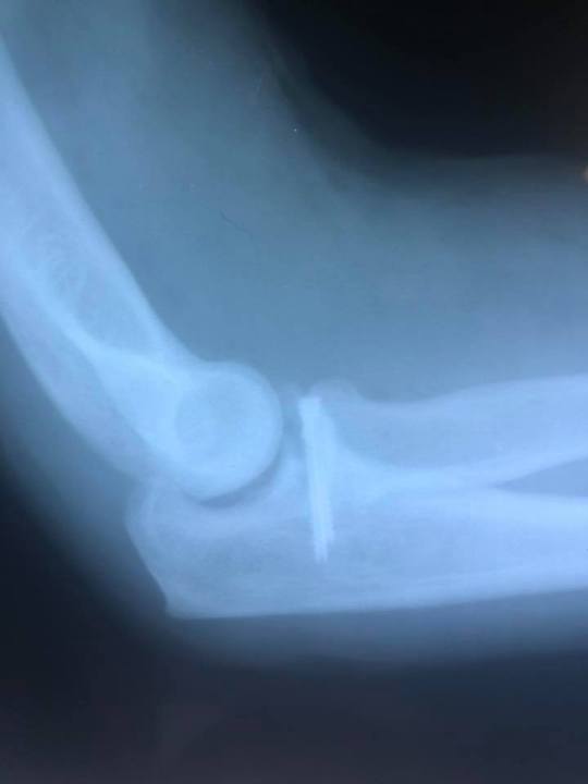
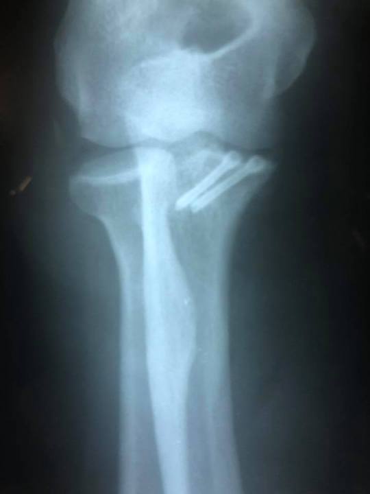
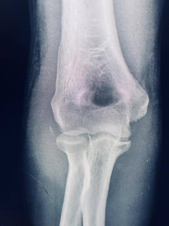
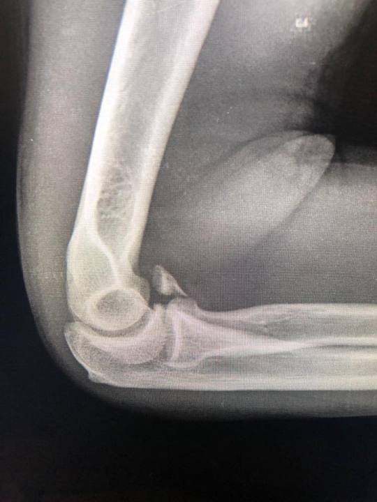
Fractura de apofisis coronoides grado III de Regan y Morrey

http://www.clinicadeartroscopia.com.mx/academia/fractura-de-apofisis-coronoides-grado-iii-de-regan-y-morrey/


Reducción abierta más fijación interna vía anterior para fractura de apofisis coronoides grado III de Regan y Morrey con tornillos de doble compresión reconstrucción articular Findmed

www.findmed.com.mx
www.drhinojosa-artroscopia.com

lunes, 11 de diciembre de 2017

Paracetamol

Diciembre 8, 2017. No. 2926
Paracetamol: actualización en su mecanismo de acción
Paracetamol: Update on its Analgesic Mechanism of Action
Christophe Mallet, Alain Eschalier and Laurence Daulhac
Pain Relief - From Analgesics to Alternative Therapies 2017
Abstract
Paracetamol is the most widely used over-the-counter medication in the world. The mechanism of action of its analgesic effect was often considered as based on the mobilization of the cyclooxygenases and more recently on serotonergic pathways. A new metabolic pathway involving the generation of an active metabolite, AM404 (N-(4- Hydroxyphenyl)-5Z,8Z,11Z,14Z-eicosatetraenamide), in the brain by the fatty acid amide hydrolase (FAAH) enzyme, was recently identified. This chapter describes experimental data that have shown the involvement of this metabolic pathway in the analgesic action of paracetamol and its relationship with the cyclooxygenase and serotonergic systems. It also explains how new targets and systems, such as the cannabinoid and vanilloid systems and the calcium channel receptor Cav3.2, play a role in the action of paracetamol. Finally, it suggests how research on the mechanism of the clinically relevant effects of this long-established analgesic could lead to new therapeutic pain strategies. Keywords: paracetamol, para-aminophenol, AM404, pain, FAAH, CB1 , TRPV1, Cav3.2, serotonin
El dolor agudo perioperatorio y el paracetamol: una visión basada en la evidencia
Dr. Alfredo Covarrubias-Gómez, Dr. José Luis González-García, Dr. José A Betancourt-Sandoval, Dr. Jonathan J Mendoza-Reyes
Rev Mex Anestesiol Volumen 36, No. 1, enero-marzo 2013
RESUMEN Antecedentes: Cada vez son más numerosos los procedimientos quirúrgicos que se realizan en México. Se prevé un incremento cercano al 20% para el próximo quinquenio. Se ha identifi cado que el 80% de los pacientes postoperados tendrá dolor, y que en el 80% de esos casos, el dolor será moderado a severo en intensidad. Para el tratamiento de dicho dolor, se han propuesto diversos abordajes terapéuticos basados en la evidencia. La piedra angular de la terapéutica postoperatoria es la analgesia regional neuroaxial epidural con anestésicos locales y opioides en combinación. No obstante, en ocasiones dicho abordaje no es posible por diversas causas. Objetivo: Fundamentar en la evidencia disponible la utilización del paracetamol en el tratamiento del dolor postoperatorio como parte un esquema analgésico estructurado. Materiales y métodos: Revisión de la literatura. Resultados: Independientemente de las controversias relacionadas con su mecanismo de acción, es posible que este fármaco tenga una actividad predominante sobre sistemas independientes (pero relacionados) a la ciclooxigenasa. Su efecto analgésico sobre placebo es signifi cativo y su efecto «ahorrador» de opioide es cercano al 50%. Se ha evidenciado que la combinación de paracetamol con AINE reduce el consumo opioide en forma signifi cativa y proporciona una mejor analgesia. Conclusiones: Se propone un esquema analgésico que incluya al opioide en combinación con paracetamol y con AINE para un control efi ciente del dolor postoperatorio. Se deben tomar en consideración las características específi cas de cada individuo y se requiere generar evidencia más robusta. Palabras clave: Manejo del dolor perioperatorio, paracetamol

International Anesthesia Research Society Annuals Meetings
USA
Like us on Facebook   Follow us on Twitter   Find us on Google+   View our videos on YouTube 
Anestesiología y Medicina del Dolor

52 664 6848905

Ventilación / Ventilation

Diciembre 9, 2017. No. 2927


Ventilación protectora y maniobras de reclutamiento pulmonar en cirugía mayor
María Camila Arango-Granados, Fredy Ariza
Rev Argent Anestesiol 2016;74:1-9 - DOI: 10.1016/j.raa.2016.05.002
Resumen
La ventilación mecánica es una intervención frecuente en el escenario quirúrgico, especialmente para procedimientos de alta complejidad y en pacientes críticamente enfermos, pero no está exenta de complicaciones. Los estudios durante los últimos años han estado encaminados a definir cuál es la estrategia ventilatoria que ofrece el mejor perfil de seguridad durante cirugía mayor. Esta revisión busca exponer cuál es la evidencia disponible sobre estrategias ventilatorias y maniobras de reclutamiento pulmonar en el paciente llevado a cirugía mayor. Para esto se realizó una búsqueda estructurada de la literatura incluida en las bases de datos MEDLINE y SciELO. Las publicaciones disponibles parecen coincidir en que el uso de volúmenes corrientes bajos reduce las complicaciones pulmonares postoperatorias en pacientes que serán sometidos a cirugía mayor. Sin embargo, existe evidencia contradictoria sobre este beneficio en cirugía de tórax. Los hallazgos sobre el uso de presión positiva al final de la espiración aún son heterogéneos. La literatura disponible indica que en cirugía de tórax, cirugía cardiovascular y cirugía traumatológica esta estrategia parece tener potenciales beneficios. En cambio, en cirugía abdominal mayor y neurocirugía existen datos contradictorios que indican la necesidad de futuras investigaciones. Aun es necesario estudiar si las maniobras de reclutamiento ofrecen reales beneficios para los pacientes que se llevan a cirugía mayor, independiente del tipo de cirugía. Finalmente, aún no es posible establecer si tanto la ventilación protectora como las maniobras de reclutamiento pulmonar realizadas en el intraoperatorio impactan en los desenlaces a largo plazo o la mortalidad.
Posición prona en pacientes con síndrome de dificultad respiratoria aguda
Prone positioning acute respiratory distress syndrome patients.
Guérin C1,2,3.
Ann Transl Med. 2017 Jul;5(14):289. doi: 10.21037/atm.2017.06.63.
Abstract
Prone position has been used in acute respiratory distress syndrome (ARDS) patients for more than 40 years in ICU. After having demonstrated its capability to significantly improve oxygenation in a large number of patients, sometimes dramatically, this procedure has been found to prevent ventilator-induced lung injury, the primary concern for the intensivists managing ARDS patients. Over the time, several trials have been done, which regularly improved and refined from each other. At the end, significant improvement in survival has been demonstrated in the most severe ARDS patients, at a threshold of 100-150 mmHg PaO2/FiO2 ratio. The effect of proning on survival cannot be predicted and seems unrelated with both severity of oxygenation impairment and oxygenation response to proning. The rate of complication is declining with the increase in centers expertise. The pressure sores are more frequent in prone and require a special attention. Prone position is a key component of lung protective mechanical ventilation and should be used as a first line therapy in association with low tidal volume and neuromuscular blocking agents in patients with severe ARDS.
KEYWORDS: Acute respiratory distress syndrome (ARDS); hypoxemia; mechanical ventilation; prone position; ventilator induced lung injury
Volumen corriente en síndrome de falla respiratoria aguda. Como mejor seleccionarlo
Tidal volume in acute respiratory distress syndrome: how best to select it.
Ann Transl Med. 2017 Jul;5(14):287. doi: 10.21037/atm.2017.06.51.
Abstract
Mechanical ventilation is the type of organ support most widely provided in the intensive care unit. However, this form of support does not constitute a cure for acute respiratory distress syndrome (ARDS), as it mainly works by buying time for the lungs to heal while contributing to the maintenance of vital gas exchange. Moreover, it can further damage the lung, leading to the development of a particular form of lunginjury named ventilator-induced lung injury (VILI). Experimental evidence accumulated over the last 30 years highlighted the factors associated with an injurious form of mechanical ventilation. The present paper illustrates the physiological effects of delivering a tidal volume to the lungs of patients with ARDS, and suggests an approach to tidal volume selection. The relationship between tidal volume and the development of VILI, the so called volotrauma, will be reviewed. The still actual suggestion of a lung-protective ventilatory strategy based on the use of low tidal volumes scaled to the predicted body weight (PBW) will be presented, together with newer strategies such as the use of airway driving pressure as a surrogate for the amount of ventilatable lung tissue or the concept of strain, i.e., the ratio between the tidal volume delivered relative to the resting condition, that is the functional residual capacity (FRC). An ultra-low tidal volume strategy with the use of extracorporeal carbon dioxide removal (ECCO2R) will be presented and discussed. Eventually, the role of other ventilator-related parameters in the generation of VILI will be considered (namely, plateau pressure, airway driving pressure, respiratory rate (RR), inspiratory flow), and the promising unifying framework of mechanical power will be presented.
KEYWORDS: Acute respiratory distress syndrome (ARDS); mechanical ventilation; tidal volume; ventilator-induced lung injury (VILI)

International Anesthesia Research Society Annuals Meetings
USA
Like us on Facebook   Follow us on Twitter   Find us on Google+   View our videos on YouTube 
Anestesiología y Medicina del Dolor

52 664 6848905

Toxicidad hepática por fármacos / Drug hepatotoxicity

Diciembre 10, 2017. No. 2928
Daño hepático inducido por drogas. ¿Conocemos todo?
Drug-induced liver injury: Do we know everything?
World J Hepatol. 2017 Apr 8;9(10):491-502. doi: 10.4254/wjh.v9.i10.491.
Abstract
Interest in drug-induced liver injury (DILI) has dramatically increased over the past decade, and it has become a hot topic for clinicians, academics, pharmaceutical companies and regulatory bodies. By investigating the current state of the art, the latest scientific findings, controversies, and guidelines, this review will attempt to answer the question: Do we know everything? Since the first descriptions of hepatotoxicity over 70 years ago, more than 1000 drugs have been identified to date, however, much of our knowledge of diagnostic and pathophysiologic principles remains unchanged. Clinically ranging from asymptomatic transaminitis and acute or chronic hepatitis, to acute liver failure, DILI remains a leading causes of emergent liver transplant. The consumption of unregulated herbal and dietary supplements has introduced new challenges in epidemiological assessment and clinician management. As such, numerous registries have been created, including the United States Drug-Induced Liver Injury Network, to further our understanding of all aspects of DILI. The launch of LiverTox and other online hepatotoxicity resources has increased our awareness of DILI. In 2013, the first guidelines for the diagnosis and management of DILI, were offered by the Practice Parameters Committee of the American College of Gastroenterology, and along with the identification of risk factors and predictors of injury, novel mechanisms of injury, refined causality assessment tools, and targeted treatment options have come to define the current state of the art, however, gaps in our knowledge still undoubtedly remain.
KEYWORDS: Acetaminophen toxicity; Acute liver failure; Cholestatic injury; Drug-induced liver injury; Hepatoxicity; Herbal-induced liver injury; Hy's law; Liver biopsy; Pharmacoepidemiology
RUCAM en daño hepático inducido por hierbas y fármacos. Actualización
RUCAM in Drug and Herb Induced Liver Injury: The Update.
Int J Mol Sci. 2015 Dec 24;17(1). pii: E14. doi: 10.3390/ijms17010014.
Abstract
RUCAM (Roussel Uclaf Causality Assessment Method) or its previous synonym CIOMS (Council for International Organizations of Medical Sciences) is a well established tool in common use to quantitatively assess causality in cases of suspected drug induced liver injury (DILI) and herb induced liver injury (HILI). Historical background and the original work confirm the use of RUCAM as single term for future cases, dismissing now the term CIOMS for reasons of simplicity and clarity. RUCAM represents a structured, standardized, validated, and hepatotoxicity specific diagnostic approach that attributes scores to individual key items, providing final quantitative gradings of causality for each suspect drug/herb in a case report. Experts from Europe and the United States had previously established in consensus meetings the first criteria of RUCAM to meet the requirements of clinicians and practitioners in care for their patients with suspected DILI and HILI. RUCAMwas completed by additional criteria and validated, assisting to establish the timely diagnosis with a high degree of certainty. In many countries and for more than two decades, physicians, regulatory agencies, case report authors, and pharmaceutical companies successfully applied RUCAM for suspected DILI and HILI. Their practical experience, emerging new data on DILI and HILI characteristics, and few ambiguous questions in domains such alcohol use and exclusions of non-drug causes led to the present update of RUCAM. The aim was to reduce interobserver and intraobserver variability, to provide accurately defined, objective core elements, and to simplify the handling of the items. We now present the update of the well accepted original RUCAM scale and recommend its use for clinical, regulatory, publication, and expert purposes to validly establish causality in cases of suspected DILI and HILI, facilitating a straightforward application and an internationally harmonized approach of causality assessment as a common basic tool.
KEYWORDS: CIOMS; RUCAM; causality assessment; dietary supplements; drug hepatotoxicity; drug induced liver injury; drugs; herb induced liver injury; herbal hepatotoxicity; herbs
Trasplante hepático en falla hepática inducida por fármacos
Liver transplantation for drug-induced acute liver failure.
Eur Rev Med Pharmacol Sci. 2017 Mar;21(1 Suppl):37-45.
Abstract
OBJECTIVES: To summarize the different clinical features of drug-induced acute liver failure, the diagnostic work-up, conservative management and the prognostic scores currently used to list patients for liver transplantation. EVIDENCE AND INFORMATION SOURCES: The current review is based on an analysis of the current literature and the caseload experience of the Authors on this topic. STATE OF THE ART: Drug-induced liver injury is the leading cause of acute liver failure in the adult population in Western countries, with a transplant-free survival rate of less than 50%. Main subtypes include paracetamol and idiosyncratic drug-induced injury, which differ in epidemiology, clinical course, prognosis and conservative management. In cases of a high likelihood of death, urgent hepatic transplantation is indicated, but the decision whether and when to put a patient with drug-induced acute liver failure on the list for urgent liver transplant is extremely difficult and requires constant interdisciplinary exchange and continuous updating of the clinical picture.
CONCLUSIONS: Intensive management should be done in a clinical tertiary referral center which has a specialized team of hepatologists and a liver transplant center.

International Anesthesia Research Society Annuals Meetings
USA
Like us on Facebook   Follow us on Twitter   Find us on Google+   View our videos on YouTube 
Anestesiología y Medicina del Dolor

52 664 6848905