lunes, 8 de mayo de 2017

El ultrasonido de hombro realizado por cirujanos ortopédicos aumenta la eficiencia en el diagnóstico de los desgarres del manguito rotador.


Shoulder ultrasonography performed by orthopedic surgeons increases efficiency in diagnosis of rotator cuff tears.

Fuente
Este artículo es originalmente publicado en:
De:
Chen YJ6,2.
2017 Apr 20;12(1):63. doi: 10.1186/s13018-017-0565-4.
Todos los derechos reservados para:
© The Author(s). 2017Open AccessThis article is distributed under the terms of the Creative Commons Attribution 4.0 International License (http://creativecommons.org/licenses/by/4.0/), which permits unrestricted use, distribution, and reproduction in any medium, provided you give appropriate credit to the original author(s) and the source, provide a link to the Creative Commons license, and indicate if changes were made. The Creative Commons Public Domain Dedication waiver http://creativecommons.org/publicdomain/zero/1.0/) applies to the data made available in this article, unless otherwise stated.
Abstract
BACKGROUND:
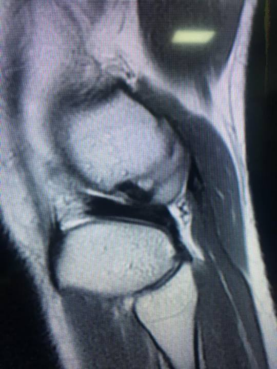
Rotator cuff tears are very common and their incidence increases with age. Shoulder ultrasonography has recently gained popularity in detecting rotator cuff tears because of its efficiency, cost-effectiveness, time-saving, and real-time nature of the procedure. Well-trained orthopedic surgeons may utilize shoulder ultrasonography to diagnose rotator cuff tears. The wait time of patients planned to have shoulder MRI (magnetic resonance imaging) to rule in rotator cuff tears may decrease after orthopedic surgeon start doing shoulder ultrasonography as a screening tool for that. Patients with rotator cuff tears may be detected earlier by ultrasonography and have expedited surgical repair. The efficacy in determination of rotator cuff tears will also increase.
CONCLUSIONS:
Office-based shoulder ultrasound examination can reduce the wait time for a shoulder MRI. The efficacy of determination of rotator cuff tears will also increase after the introduction of shoulder ultrasonography.
KEYWORDS:
Diagnosis; Efficiency; Rotator cuff; Shoulder; Ultrasonography
Resumen

ANTECEDENTES:
Los desgarres del manguito rotador son muy comunes y su incidencia aumenta con la edad. La ultrasonografía de hombro ha ganado recientemente popularidad en la detección de los desgarres del manguito rotador debido a su eficiencia, rentabilidad, ahorro de tiempo, y en tiempo real la naturaleza del procedimiento. Los cirujanos ortopédicos bien entrenados pueden utilizar la ecografía o ultrasonido del hombro para diagnosticar los desgarros del manguito rotador. El tiempo de espera de los pacientes proyectados para someterse a RM del hombro (resonancia magnética) para descartar los desgarres del manguito rotador puede disminuir después de que el cirujano ortopédico comience a hacer ultrasonografía en el hombro como una herramienta de detección para eso. Los pacientes con desgarramiento del manguito rotador pueden ser detectados antes por ultrasonografía y han acelerado la reparación quirúrgica. La eficacia en la determinación de los desgarres del manguito rotador también aumentará.

CONCLUSIONES:
El examen de ultrasonido de hombro basado en la oficina puede reducir el tiempo de espera para una RM de hombro. La eficacia de la determinación de los desgarres del manguito rotador también aumentará después de la introducción de la ecografía del hombro.

PALABRAS CLAVE:
Diagnóstico; Eficiencia; Manguito rotador; Hombro; Ultrasonografía
PMID:  28427416   PMCID:  
  DOI:  

Libro sobre pediatría / Book on pediatrics

Mayo 7, 2017. No. 2682


Cirugía pediátrica y neonatal
Pediatric and Neonatal Surgery
Edited by Joanne Baerg, ISBN 978-953-51-3136-6, Print ISBN 978-953-51-3135-9, 168 pages, Publisher: InTech, Chapters published May 03, 2017 under CC BY 3.0 license
Edited Volume
Neonatal and Pediatric Surgery is a broad field with many challenges. The aim of this short book is to provide the reader with several informative chapters in the field of neonatal and pediatric surgery. Each chapter provides details on a specific area of this changing field. The scope of this book focuses on a few areas that are rare and challenging. For example, it covers preoperative and postoperative care of neonates. Important anesthesia considerations, including anesthesia for neonates and regional anesthesia, are discussed. A unique chapter on neonatal tumors is presented. The book provides an overview of the recent recommendations for care of infants and children that undergo cardiac surgery. The challenging aspects of caustic ingestion are explained. Each chapter stands alone as a detailed source of information for the reader. This book brings updated information with structured headings that will allow the reader to remain focused as the material is reviewed.

Like us on Facebook   Follow us on Twitter   Find us on Google+   View our videos on YouTube 
Anestesiología y Medicina del Dolor

52 664 6848905

Trasplante hepático / Liver transplant

Mayo 8, 2017. No. 2683


Hemorragia masiva en trasplante hepático: Consecuencias, predicción y manejo
Massive haemorrhage in liver transplantation: Consequences, prediction and management.
World J Transplant. 2016 Jun 24;6(2):291-305. doi: 10.5500/wjt.v6.i2.291.
Abstract
From its inception the success of liver transplantation has been associated with massive blood loss. Massive transfusion is classically defined as > 10 units of red blood cells within 24 h, but describing transfusion rates over a shorter period of time may reduce the potential for survival bias. Both massive haemorrhage and transfusion are associated with increased risk of mortality and morbidity (need for dialysis/surgical site infection) following liver transplantation although causality is difficult to prove due to the observational design of most trials. The blood loss associated with liver transplantation is multifactorial. Portal hypertension secondary to cirrhosis results in extensive collateral circulation, which can bleed during hepatectomy particular if portal pressures are increased. Avoiding volume loading and maintenance of a low central venous pressure together with the use of vasopressors have been shown to reduce blood loss and transfusion during liver transplantation, but may increase the risk of renal impairment post-operatively. Coagulation defects may be present pre-transplant, but haemostasis is often re-balanced due to a deficit in both pro- and anti-coagulation factors. Further derangement of haemostasis may develop in the anhepatic and neohepatic phases due to absent hepatic metabolic function, hyperfibrinolysis and platelet sequestration in the donor liver. Point-of-care tests of coagulation such as the viscoelastic tests rotation thromboelastometry/thromboelastometry allow and more accurate and rapid assessment of these derangements in coagulation and guide the use of factor replacement and antifibrinolytics. Transfusion protocols guided by these tests have been shown to reduce transfusion rates compared with conventional coagulation tests, but have not shown improvements in mortality or morbidity. Pre-operative factors associated with massive transfusion include previous surgery, re-do transplantation, the aetiology and severity of liver disease. Intra-operatively the use of piggy-back technique and avoiding veno-veno bypass has been shown to reduced blood loss.
KEYWORDS: Coagulopathy; Liver transplantation; Massive transfusion

Complicaciones trombóticas perioperatorias en trasplante hepático
Perioperative thrombotic complications in liver transplantation.
World J Gastroenterol. 2015 Jul 14;21(26):8004-13. doi: 10.3748/wjg.v21.i26.8004.
Abstract
Although the perioperative bleeding complications and the major side effects of blood transfusion have always been the primary concern in liver transplantation (OLT), the possible cohesion of an underestimated intrinsic hypercoagulative state during and after the transplant procedure may pose a major threat to both patient and graft survival. Thromboembolism during OLT is characterized not only by a complex aetiology, but also by unpredictable onset and evolution of the disease. The initiation of a procoagulant process may be triggered by various factors, such as inflammation, venous stasis, ischemia-reperfusion injury, vascular clamping, anatomical and technical abnormalities, genetic factors, deficiency of profibrinolytic activity, and platelet activation. The involvement of the arterial system, intracardiac thrombosis, pulmonary emboli, portal vein thrombosis, and deep vein thrombosis, are among the most serious thrombotic events in the perioperative period. The rapid detection of occlusive vascular events is of paramount importance as it heavily influences the prognosis, particularly when these events occur intraoperatively or early after OLT. Regardless of the lack of studies and guidelines on anticoagulant prophylaxis in this setting, many institutions recommend such an approach especially in the subset of patients at high risk. However, the decision of when, how and in what doses to use the various chemical anticoagulants is still a difficult task, since there is no common consensus, even for high-risk cases. The risk of postoperative thromboembolism causing severe hemodynamic events, or even loss of graft function, must be weighed and compared with the risk of an important bleeding. In this article we briefly review the risk factors and the possible predictors of major thrombotic complications occurring in the perioperative period, as well as their incidence and clinical features. Moreover, the indications to pharmacological prophylaxis and the current treatment strategies are also summarized.
KEYWORDS: Hepatic artery occlusion; Liver transplantation; Postoperative complications; Pulmonary emboli; Thromboembolic phenomena; Vascular 

Falla renal aguda después de trasplante hepático. Revisión sistemática de modelos predictivos publicados
Acute kidney injury following liver transplantation: a systematic review of published predictive models.
Anaesth Intensive Care. 2016 Mar;44(2):251-61.
Abstract
Acute kidney injury is a frequent postoperative complication amongst liver transplant recipients and is associated with increased morbidity and mortality. This systematic review analysed the existing predictive models, in order to solidify current understanding. Articles were selected for inclusion if they described the primary development of a clinical prediction model (either an algorithm or risk score) to predict AKI post liver transplantation. The database search yielded a total of seven studies describing the primary development of a prediction model or risk score for the development of AKI following liver transplantation. The models span thirteen years of clinical research and highlight a gradual change in the definitions of AKI, emphasising the need to employ standardised definitions for subsequent studies. Collectively, the models identify a diverse range of predictive factors with several common trends. They emphasise the impact of preoperative renal dysfunction, liver disease severity and aetiology, metabolic risk factors as well as intraoperative variables including measures of haemodynamic instability and graft quality. Although several of the models address postoperative parameters, their utility in predictive modelling seems to be of questionable relevance. The common risk factors identified within this systematic review provide a minimum list of variables, which future studies should address. Research in this area would benefit from prospective, multi-site studies with larger cohorts as well as the subsequent internal and external validation of predictive models. Ultimately, the ability to identify patients at high risk of post-transplant AKI may enable early intervention and perhaps prevention.
KEYWORDS: acute kidney injury; liver transplantation; prediction model; risk score

Like us on Facebook   Follow us on Twitter   Find us on Google+   View our videos on YouTube 
Anestesiología y Medicina del Dolor

52 664 6848905

Lesión en Asa de Balde meniscal


Lesión en Asa de Balde meniscal                                                                                                                                                       
Imagen de doble cruzado posterior correspondiente a lesión en Asa de Balde meniscal se realizo sutura con fast fix 360 más reconstrucción de ligamento cruzado anterior autologo femenino 21 años

Grupo Cirugía y Reconstrucción Articular 

Fractura de Jones


Fractura de Jones manejado con tornillo canulado doble compresión por mínima invasión, fractura consolidada al 100%
http://www.clinicadeartroscopia.com.mx/academia/fractura-de-jones/