sábado, 30 de abril de 2011

Taking Aim at Melanoma


Taking Aim at Melanoma

Understanding oncogenesis at the molecular level offers the prospect of tailoring treatments much more precisely for patients with advanced cases of this deadliest of skin cancers.

Pep Karsten / fstop / Corbis
They’re lawyers and receptionists, philanthropists and film editors. Some are retired, some just starting families. What they have in common is metastatic melanoma, a cancer that will likely claim their lives in a matter of months. Standard treatments—including removal of tumors and chemotherapy—have failed to halt their advancing disease. But these patients have not given up. All have volunteered to take part in clinical trials designed to test novel therapies that could help rein in their tumor growth and possibly buy them some time. And thanks to two promising new approaches, these patients—and others like them—have reason to be hopeful.
The new treatments are more carefully aimed than the massive but indiscriminate hammer blow dealt by standard chemotherapeutics, which have always disappointed in the treatment of melanoma. One approach takes advantage of drugs that have been designed specifically to take down the mutant protein that is inappropriately activated in more than half of all patients with melanoma. Another is a form of immunotherapy that activates T cells that can recognize and attack the tumors. Both have shown an ability to improve the lives of patients with metastatic melanoma, and both are likely to gain FDA approval this year. The results offer hope that someday we may be able to overcome this disease, for which no effective treatments currently exist.
Identifying a culprit gene
Melanoma is the deadliest form of skin cancer. When the disease is caught early, it can be easily treated. But once the cancer spreads, the prognosis is bleak. The standard form of chemotherapy involves treatment with dacarbazine, the only drug approved for treatment of stage IV metastatic melanoma. Dacarbazine interferes with cell growth by chemically modifying DNA, and it is no miracle drug. Only 10 percent of melanoma patients show any response to the drug, and the treatment ultimately has no effect on overall survival.
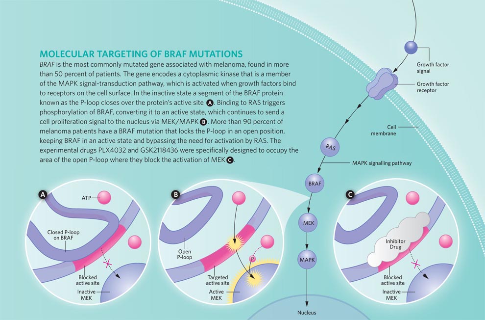
Drugs like dacarbazine are indiscriminate: they batter all cells, whether normal or cancerous. When taking aim at cancer cells, it helps to have a more precisely defined target. In 2002, investigators led by Chris Marshall at the Institute of Cancer Research in London identified such a target. The researchers discovered mutations in a protein kinase called BRAF in more than half of the melanomas they examined.1 These mutations inappropriately activate the kinase, which then turns on a second kinase called MEK. This activational cascade—part of the well-studied MAP kinase signaling pathway—ultimately turns on a suite of genes that mediates cell division. (See “On the MAP,”The Scientist, June 2009.)
The discovery that BRAF activation is a key feature of many melanomas raised the possibility that investigators would be able to identify compounds that would specifically target the aberrant signals that drive this form of cancer. In the years following the identification of BRAF mutations in melanoma, we turned our attentions to sorafenib—a drug that was being tested (and ultimately approved) for the treatment of renal cancer. Sorafenib is a potent inhibitor of the receptors that bind to the angiogenesis-promoting hormone VEGF. It also blocks the activities of a handful of protein kinases, including BRAF.
Unfortunately, we found that sorafenib is a poor inhibitor of BRAF in patients. In 2002 and 2004, I led the clinical trials to assess its ability to act as a single-agent therapeutic for patients with melanoma. In Phase I trials, the maximal dose that could be tolerated by patients was determined, and in Phase II we gave patients as much sorafenib as they could safely handle. But even at the maximal doses, sorafenib showed little to no clinical efficacy in melanoma patients. And biopsies revealed that the drug was not blocking BRAF activity effectively.2,3
click to enlarge
Infographic: Molecular Targeting of Braf Mutations
View full size JPG (120 KB) | PDF (560 KB)
Lucy Reading-Ikkanda
But we still had high hopes for the targeted approach. Sorafenib was in effect a part-time, accidental BRAF inhibitor—and not a very good one in the end. So the failure of those early trials left the door open for more potent, selective, “professional” BRAF inhibitors to come along. Drugs that were designed specifically to shut down overactive BRAF might be more effective at curbing tumor growth and improving patients’ lives. And with the targeted BRAF inhibitors PLX4032 and GSK2118436, our hopes were realized.4
Attacking BRAF
BRAF is a protein of 766 amino acids. The most common mutation found in melanoma changes the valine (V) at position 600 to a glutamic acid (E). This alteration—dubbed V600E in shorthand—accounts for 90 percent of the mutations in tumor samples analyzed. The next most common mutation, present in a handful of melanomas, changes that same valine to a lysine (V600K). A smattering of other mutation also occur in and around that amino acid hot spot.1
Although targeted BRAF inhibitors have shown promise for malignant melanoma, coming up with more effective combination therapies tops the agenda.
All these mutations function to lock BRAF in its active state. When the protein is inactive, a segment called the P-loop covers the site where ATP binds. When BRAF is activated, the P-loop swings out, exposing this site and allowing the kinase to phosphorylate its target protein, MEK.4 Structural studies, particularly of the V600E form of BRAF, show that mutations repel the P-loop, keeping it from shutting down the kinase activity. (See illustration above.)
The first targeted BRAF inhibitor was designed specifically to recognize and shut down the V600E form of the protein. Developed by scientists at a small biotech company called Plexxikon, the compound—PLX4032—binds to BRAF in its active conformation and prevents it from interacting with its target protein MEK.4 In blocking the activation of MEK, PLX4032 arrests progression into the G1 phase of the cell cycle, halting proliferation. And it also triggers apoptosis—in vitro, in animal models, and in the tumors of patients enrolled in Phase I/II clinical trials. 5
Next to bat
GSK2118436 is a drug developed by the pharmaceutical company GlaxoSmithKline (GSK). Like PLX4032, the GSK compound is a highly selective inhibitor of mutant forms of BRAF. Both bind to the same region of the protein and help hold it in its inactive conformation. But the GSK compound is even more potent than PLX4032: it cuts BRAF’s activity in half at concentrations less than 1 nM. Clinical trials for this potent inhibitor have accepted patients who have any form of the V600 mutant BRAF proteins. Most of those enrolled have the V600E form of mutant BRAF, but a few have the V600K or V600G (valine to glycine) mutation.
Although GSK2118436 has been administered to fewer patients than PLX4032, both drugs have shown similar efficacies in terms of tumor shrinkage. At the maximal dosages, about 90 percent of patients with BRAF mutations show some degree of tumor regression; 60 to 70 percent of patients had tumors that shrank by one-third or more.6 The trials have not gone on long enough to assess either drug’s effect on overall survival, but compared to treatment with dacarbazine, PLX4032 appears to triple the length of time patients have before the disease starts to worsen—from two months with dacarbazine to six months with the new drug.
The GSK compound had the added benefit of actually reducing the size of some of the small brain metastases seen in a subgroup of the patients enrolled in that trial.5 This response was particularly welcome because half of all patients with malignant melanoma will develop brain tumors, which respond particularly poorly to treatment. Once melanoma spreads to the brain, survival can be a matter of weeks, particularly for those with large, symptomatic metastases. So GSK2118436’s ability to shrink brain tumors is being actively pursued.
The trials have been an emotional roller coaster for the patients receiving the drugs—and for the researchers and medical personnel developing and administering them. [See this three-part series, “Target Cancer” featuring Keith Flaherty (The New York Times, Feb 22-24, 2010).] And the benefits seen in the Phase II clinical trials should garner targeted BRAF inhibitors FDA approval sometime in 2011. But the news is not all good. A response rate of 90 percent means that 10 percent of the patients enrolled in the PLX and GSK trials are not helped by these targeted drugs. Their tumors continued to grow during the course of the treatment. In addition, a large percentage of those whose tumors shrank during the first months of treatment will develop drug resistance. Investigators are still exploring what the mechanism of this resistance might be. What they have determined so far is that patients do not seem to accrue any new, reactivating mutations in BRAF itself. Instead, some tumors show mutations that enhance the activity of other kinases in the MAP kinase pathway to which BRAF belongs.7 Other tumors accumulate mutations that activate different signaling pathways which mediate cell proliferation or survival, including that of another oncogenic kinase called phosphoinositide 3-kinase (PI 3-kinase).8
Getting a handle on how tumors can circumvent BRAF inhibitors should give us a better idea of how to attack melanomas that become resistant to these promising new agents. The knowledge could also point the way toward drugs that could be effectively combined with BRAF-targeted inhibitors as a frontline form of therapy for metastatic melanoma to prevent the emergence of these resistance mechanisms. BRAF inhibitors could be given along with targeted inhibitors of other oncogenic kinases, including PI 3-kinase. Another approach would be to combine targeted BRAF inhibitors with an entirely different type of treatment, such as immunotherapy.9
The long haul
Even if targeted molecular therapies like PLX4032 or the GSK compound were 100 percent effective, only half of all patients with melanoma have tumors with BRAF mutations. For the other half, we need to come up with some viable alternatives. Some of the patients have other mutations that might serve the same function as BRAF. For those patients, similar treatment strategies are being developed. But for any patient with melanoma, immunotherapy provides an option that does not appear to depend on specific mutations in the tumor.
click to enlarge
Infographic: Boosting T Cell Activation
View full size JPG (205 KB) | PDF (171 KB)
Lucy Reading-Ikkanda
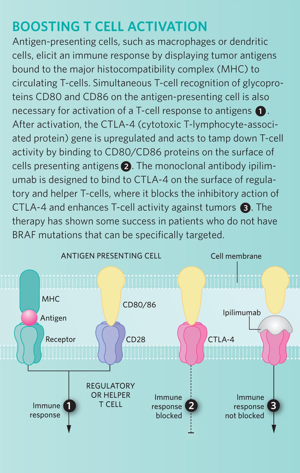
Perhaps the most directed approach would be to prime the immune system to recognize antigens that are specific to melanomas—or at least to the melanocytes that give rise to this cancer. In the mid-1990s, investigators identified a protein that could potentially serve as a target antigen: a membrane-bound glycoprotein called gp100. This protein is present on the surface of cells in the melanocyte lineage, as well as cells of the majority of both primary and metastatic melanomas.
By formulating a vaccine that consists of a small fragment of gp100, researchers hoped they could convince the patient’s immune system to attack tumor cells bearing this melanoma-specific antigen. Unfortunately, the gp100 peptide vaccine has never shown much clinical benefit when given on its own. But animal models and some small clinical trials suggest that when combined with an agent that can also stimulate a patient’s T cells, gp100 could help target immune cells to the tumors that are to be destroyed. One of these T-cell–boosting drugs is ipilimumab.
Ipilimumab (made by Bristol-Myers Squibb) is a monoclonal antibody that recognizes the CTLA4 protein on the surface of regulatory and helper T cells.10 CTLA4 inhibits T-cell activation and helps to promote immunologic tolerance, so blocking this protein should stimulate an immune response and help prime T cells to attack tumors bearing antigenic target proteins.
The results of the first large phase III trial for ipilimumab look promising. In this trial, investigators compared patients who received either a combination of the gp100 vaccine plus ipilimumab to those who received either of the treatments on its own. In this case, the vaccine-only patients served as a “control arm” of the study, since gp100 has not been found to be effective in treating melanoma when administered as a single agent. The results confirmed what had been seen in smaller, nonrandomized studies in which everyone received the drug: about 10 percent of the patients taking ipilimumab—either with or without the peptide vaccine—experienced tumor shrinkage of one-third or more.11 As yet, we do not know why these patients responded to the drug, while others did not. One goal for the future would be to identify what makes these responders unique so that we can target this therapy to the patient population most likely to benefit.
Even though 90 percent of the patients showed no shrinkage, it does not necessarily mean that the drug did them no good. For some of these patients, ipilimumab (again, taken alone or in conjunction with the peptide vaccine) has halted disease progression: their tumors have remained the same size for years—and they are still alive to tell about it. Such a response is unusual for patients with malignant melanoma, a cancer that generally progresses quite rapidly. And among the 10 percent of patients who initially responded to the drug, most continue to benefit. In the randomized trial it was clear that a larger percentage of patients lived for two years after receiving ipilimumab, and from earlier trials some who have been responding for 10 years and counting.
Another large phase III trial—comparing ipilimumab plus dacarbazine to dacarbazine alone—has been going on for several years and we hope the results of that study will corroborate the life-prolonging effects we’ve already seen. But even without that trial, many of us believe that the FDA will look favorably on the results reported thus far, because no other drug tested to date has ever enhanced survival in a phase III trial for malignant melanoma.
More one-two punches
Although targeted BRAF inhibitors have shown promise, none of us is satisfied with what single-agent therapies have accomplished thus far. So for malignant melanoma, coming up with more effective combination therapies tops the agenda. The low-hanging fruit would involve using drugs already developed for targeting other kinases or other pathways in which aberrant activity contributes to the development of melanoma, such as members of the PI 3-kinase pathway.
At the same time, oncologists are migrating toward mixing different types of therapies, such as targeted BRAF inhibitors plus immunotherapy. Our group at Massachusetts General Hospital has produced data supporting the idea that combining those approaches would likely benefit our patients. We have found that melanoma cells exposed to BRAF inhibitors become more densely decorated with surface antigens such as gp100. In the absence of treatment, these cancer cells somehow suppress expression of their melanocyte-specific antigens, which presumably helps them to escape immune surveillance. And BRAF inhibitors seem to reverse this suppression. Working with melanoma biopsies taken before and after therapy with BRAF inhibitors, we have found that treated tumor cells display more melanocyte- specific antigens and are more easily recognized by T cells harvested from the patient.9
Of course, convincing pharmaceutical companies to make their drugs available for such combination trials can in itself be challenging. First, the specificity of each treatment could mean that any combination would further fractionate the population of patients for whom the treatment would work. For example, more than half of patients with melanoma have a mutation in BRAF. And a smaller percentage of those will have a BRAF mutation combined with a companion mutation in another kinase for which an inhibitor is available or can be developed. Drug companies can be loath to move forward with therapies for which the market promises to be small.
Pharmaceutical companies also shy away from coadministering drugs because of the fear that a drug combination could produce a unique toxicity—something that would not be an issue with either drug alone. Such a reaction could kill a compound that’s still in development—even if that drug would be perfectly safe and perhaps effective when taken as a single agent.
Cancer is a complex disease, and developing effective treatments is more complex still. For the benefit of patients, my colleagues and I contend that the whole drug development industry needs to recognize this complexity and incentives need to be provided to companies that are willing to take on the challenge of formulating new therapies—and combinations of therapies—to tackle this disease. The science is out there to support these approaches. Now, we as physicians need to show as much determination as our patients in our ongoing search for the drugs—or combinations of drugs—that will allow people with malignant melanoma to return home and go on living their lives.
Andrzej Tokarski / ISTOCKPHOTO.COM
A DIET OF PILLS
There comes a point in any trial when investigators agree to pause and take stock of the situation. In Phase I trials, for example, we typically stop to determine whether we’re seeing any side effects that would warrant discontinuing the trial—or whether we’re seeing any effects that would suggest we move forward and expand the number of patients receiving the drug. In the case of PLX4032, developed at a small California biotech company called Plexxikon, we ran into a different problem: we hit a plateau in terms of bioavailability. In other words, we could increase the dosage of drug the patients were receiving, but we couldn’t get their blood concentrations high enough.
On the one hand, we could have opted to push forward, persuading our patients to swallow even more of the poorly absorbed pills. But there are drawbacks to that approach. First, overloading people’s digestive systems with drugs can produce local irritation, resulting in diarrhea and the like. Although these conditions are not life-threatening, they can make it difficult for patients to tolerate the therapy—not a good outcome for any new drug. And even if the megadoses don’t cause intestinal distress, a high pill count can cause problems down the road. Patients in Phase I trials are a highly motivated cohort, willing to serve as guinea pigs on drugs that have never before been tested in humans. But what if patients in later trials are not as willing to do whatever it takes? If your drug only works at the highest dose, but your patients are not willing to swallow more than 30 pills a day to achieve that dose, then the trial will fail to show efficacy, and a potentially useful drug could be permanently shelved.
Instead, we voted to pause—to wait as long as it would take to derive a better formulation. If that formulation could not be found, we would consider walking away, trying to find another agent that could achieve the concentrations that would be needed to really take down the mutant BRAF. Fortunately, Plexxikon and Roche (the company’s new partner in developing PLX4032) were willing and able to rework the drug into a form that boosted blood concentrations higher. Even then, we saw no significant side effects, so we increased the dosage—to the point that people were taking 14 capsules, twice a day. That was a steady diet of pills.
Since then, the drug has been made even more concentrated, so fewer, smaller capsules will yield the same effect. It’s important to remember that we are working with people here—people we wish to help. So we have to respect their limits. In doing so, we not only benefit the patients in our trial, but all patients who will ultimately benefit from receiving an effective treatment in the future.
Editor's Note (4th April): Just prior to the publication of this article, the FDA approved ipilimumab for use in advanced melanoma patients. It was shown to lengthen the life spans of 20-30% of advanced melanoma patients participating in a clinical trial of the drug. Some patients lived several years after their initial diagnosis.
F1000 Member Keith T. Flaherty is Director of Developmental Therapeutics at Massachusetts General Hospital Cancer Center in Boston.


AlphaMed Press, publisher of THE ONCOLOGIST® and THE ONCOLOGIST COMMUNITYSM website, grants The Scientist permission to provide this video link to Keith T. Flaherty’s lecture, "BRAF: the first prevalent oncogene target to show single-agent responsiveness," at THE ONCOLOGIST COMMUNITY.

 
1. H. Davies et al.,“Mutations of the BRAF gene in human cancers,” Nature, 417:949-54, 2002. Free F1000 Evaluation
2. K.T. Flaherty et al.,“Phase I/II, pharmacokinetic and pharmacodynamic trial of BAY 43-9006 alone in patients with metastatic melanoma,” J Clin Oncol, 23(16s): abstract 3037, 2005.
3. T. Eisen et al., “Sorafenib in advanced melanoma: a Phase II randomised discontinuation trial analysis,” Br J Cancer, 95:581-86, 2006.
4. G. Bollag et al., “Clinical efficacy of a RAF inhibitor needs broad target blockade in BRAF-mutant melanoma,”Nature, 467: 596-99, 2010. Free F1000 Evaluation
5. R. A. Kefford et al., “Phase I/II study of GSK2118436, a selective inhibitor of oncogenic mutant BRAF kinase, in patients with metastatic melanoma and other solid tumors,” J Clin Oncol, 28(15s): abstract 8503, 2010.
6. K. T. Flaherty et al., “Inhibition of mutated, activated BRAF in metastatic melanoma,” N Engl J Med, 363:809-19, 2010. Free F1000 Evaluation
7. C.M. Johannessen et al., “COT drives resistance to RAF inhibition through MAP kinase pathway reactivation,”Nature, 468:968-72, 2010. Free F1000 Evaluation
8. R. Nazarian et al., “Melanomas acquire resistance to B-RAF(V600E) inhibition by RTK or N-RAS upregulation,”Nature, 468:973-77, 2010.
9. A. Boni et al., “Selective BRAFV600E inhibition enhances T-cell recognition of melanoma without affecting lymphocyte function,” Cancer Res, 70:5213-19, 2010. Free F1000 Evaluation
10. J.S. Weber et al., “Phase I/II study of ipilimumab for patients with metastatic melanoma,” J Clin Oncol,26:5950-56, 2008.
11. F.S. Hodi et al., Improved survival with ipilimumab in patients with metastatic melanoma, N Engl J Med,363:711-23, 2010. Free F1000 Evaluation

An Aspirin for your Cancer?


An Aspirin for your Cancer?

Can tumors—which can originate from, and often resemble, chronically inflamed tissue—be curtailed using familiar anti-inflammatory agents, without their side effects?

Colin Anderson / Gettyimages
What if taking aspirin could reduce your risk of cancer? Researchers have debated the relationship between inflammation and cancer for many years, but recent studies have reignited the discussion with evidence that taking aspirin daily for 5 years or longer can protect against death from colorectal and other solid cancers. If this observation indeed holds true, and aspirin can stave off cancer or reduce the risk of recurrence, this familiar, age-old drug could offer a tantalizingly simple treatment.

Unfortunately, aspirin and other nonsteroidal anti-inflammatory drugs (NSAIDs) are not without problematic side effects, increasing the risks of liver toxicity and bleeding in the stomach and brain when taken over extended periods of time. Researchers who have been studying the molecular pathways at the intersection of cancer and inflammation hope their findings may lead to more selective ways of reducing inflammation, eliminating or minimizing aspirin’s negative effects without sacrificing its benefits.
When Peter Rothwell at John Radcliffe Hospital in Headington, Oxfordshire, and colleagues analyzed individual patient data from eight randomized trials in which patients took a daily aspirin for prevention of cardiovascular diseases, they noticed the aspirin takers had a lower incidence of death from cancer than those who didn’t take the drug.1 Earlier studies had shown that daily use of aspirin and other NSAIDs over extended periods reduced the risk of colorectal cancer or polyp recurrence, but no clear evidence was previously available, at least in humans, that aspirin might also reduce the risk of other cancers. In the new study, the benefit of aspirin use was apparent after at least five years of treatment. In trials in which the patients had been taking aspirin for more than 7.5 years, the 20-year risk of cancer death (from the initiation of the trials) was reduced by approximately 30 percent for all solid cancers and by 60 percent for gastrointestinal cancers. For lung and esophageal cancer, the benefit was confined to subtypes of those cancers that originated in glandular tissue (adenocarcinomas). For colorectal cancer, the effect was high for cancer in the proximal colon but not in the distal colon.
These data clearly point to the importance of anti-inflammatory drugs in preventing the initiation and progression of both gastrointestinal and other solid organ cancers (including lung and prostate), and suggest that inflammation may be an underlying cause of cancer even in tumor types that had not been traditionally thought to originate within chronically inflamed tissues.
Inflammation and cancer genes
Although the role of inflammation in favoring carcinogenesis has generated much interest in the last 10–15 years, the Greek physician Claudius Galenus observed some similarity between cancer and inflammation almost 2 thousand years ago. Galenus originally used Hippocrates’s term “cancer” specifically to describe certain inflammatory tumors of the breast in which superficial veins appeared swollen and radiated, somewhat like the claws of a crab. Later the name was extended to include all malignant and infiltrating growths. In 1863 Rudolf Virchow noted white blood cells or leukocytes in neoplastic tissues and made a connection between inflammation and cancer. He suggested that the “lymphoreticular infiltrate” reflected the origin of cancer at sites of chronic inflammation. A seminal observation was made more than a century later, when Harold Dvorak of Harvard University noted that inflammation and cancer share some basic developmental mechanisms (angiogenesis) and tissue-infiltrating cells (lymphocytes, macrophages, and mast cells), and that tumors act like “wounds that do not heal.”
Chronic inflammation can affect all phases of carcinogenesis, from favoring the initial genetic alterations that drive cancer formation, to acting as a tumor promoter by establishing conditions in the surrounding tissues that allow the tumor to progress and metastasize, and even triggering immunosuppressive mechanisms that prevent an effective immune response against the tumors.
In 2004 Robert Bass Jr. at the The University of Texas M. D. Anderson Cancer Center and colleagues showed for the first time that the cancer gene RAS also plays an important role in inflammation.2 Recent studies furthered this research, revealing that such genes often have dual roles. Many genes that are known to play a role in cancer when they are abnormally activated—the oncogenes RAS, RET, BRAF, SRC, and MYC—appear to play a role in inflammation as well. These genes turn on the inflammatory pathway within the cell, as well as activating inflammation outside the cell, by recruiting and initiate inflammatory cells that create an environment which reduces anticancer immune cell defenses.3 Interestingly, by way of epigenetic changes, continued activation or overexpression of oncogenes may not always be required for maintaining this pro-inflammatory loop: transient activation of the SRC oncoprotein induces an epigenetic switch that uses microRNA regulation to stably maintain the production of IL-6, a key inflammatory cytokine.4 Severe DNA damage, such as double-stranded breaks, activates the ataxia telangiectasia-mutated (ATM) enzyme, a kinase that repairs DNA but also turns on the secretion of pro-inflammatory factors. These same factors go on to create conditions that promote an oncogenic growth of cells with double-strand breaks, thereby maintaining the production of proinflammatory factors through a positive feedback loop.5
click to enlarge
Infographic: Where Cancer and Inflammation Intersect
View full size JPG (207 KB) | PDF (123 KB) 
Lucy Reading-Ikkanda
In recent years, researchers have begun to appreciate the role that a tumor’s environment plays in its growth and survival. Indeed, inflammation of the tissue surrounding a tumor can hasten the oncogenic process by directly promoting genetic instability and favoring or inducing gene mutations. Reactive oxygen and nitrogen species (ROS and RNS), which are abundant during inflammation, can induce DNA mutations, epigenetic alterations, and posttranslational modifications of proteins that control the cell cycle or survival. In particular, ROS and RNS have been shown to reduce the expression and enzymatic activity of DNA mismatch repair (MMR) proteins such as mutL homolog 1 (MLH1) and mutS homologs 2 and 6 (MSH2 and MSH6), resulting in increased genetic instability.6Researchers also noted that a protein called AID, which actively mutates DNA during B-cell maturation, was abnormally turned on in inflamed epithelial cells—an effect linked to colorectal cancer. These mechanisms suggest that genomic instability, epigenetic changes, and functional protein modifications are involved in the early events of inflammation-induced cancer initiation.
Crisscrossing inflammatory and cancer pathways
A number of innate immune receptors, like the numerous members of the Toll-like receptor (TLR) family and other pattern-recognition receptors (PRRs), activate inflammatory signals within the cell when they encounter signs of bacterial or viral infection such as nucleic acids and pieces of bacterial cell wall or flagellum. Recent evidence has shown that the pathways that are activated by these innate immune receptors also activate functions—such as cell proliferation, differentiation, and cell death—which can predispose a cell to cancer.7
These receptors activate the cell’s first-responder mechanism, which turns on major transcription factors that quickly activate genes to prepare cells for the onslaught of damage. The transcription factors, NF-κB, STAT3, and the adaptor protein MyD88—each of them key to the innate inflammatory response—are also proving to be essential in certain kinds of cancers.
NF-κB was first discovered by David Baltimore in 1986 as a protein involved in immunity and was subsequently shown to trigger innate inflammation.8 Today, research is beginning to indicate that its expression is also required for oncogenesis in many tissues.9 NF-κB activates genes involved in regulating the cell cycle, angiogenesis, and cell survival, as well as genes encoding pro-inflammatory cytokines, chemokines, and proteases—processes that can play major roles in cancer initiation and progression when not properly kept in check.
STAT3 is another major transcription factor involved in immunity and inflammation. It is also overexpressed and phosphorylated in most types of cancer. STAT3 contributes to tumor cells’ survival, proliferation, and dissemination by controlling the expression of several cell-cycle genes and of the proto-oncogene c-MYC. STAT3 also favors survival of malignant cells by preventing apoptosis, in part by transcriptional downregulation of p53, and by controlling pro-angiogenic and metastatic factors such as VEGF and metalloproteases.9
In skin cancers, expression of STAT3 in epithelial cells is required for the initiation of cancer formation, suggesting that inflammation is a necessary component of carcinogenesis in this tissue. Paradoxically, when turned on in cancer cells, STAT3 activates expression of chemokines that promote inflammation and immunity. However, when it is activated in immune cells, the same pathway initiates anti-inflammatory or immunosuppressive signals that shield the tumor from immune attack. These contrasting activities of STAT3 favor tumor growth because the activated immune cells that are recruited to the site of inflammation provide factors for angiogenesis and rebuilding of the extracellular matrix, both of which favor tumor growth, while the strong inflammatory and immune responses that are associated with antitumor and anti-angiogenic effects are prevented.9
Both NF-κB and STAT3 are activated and regulated in their activities by a number of mechanisms and regulatory molecules. One such mechanism is controlled by the signaling adaptor protein MyD88, which is required for the downstream signaling of most of the Toll-like receptors (TLRs) and the interleukin-1 (IL-1) family of pro-inflammatory cytokines. MyD88 is central for the activation of NF-κB and some of the other most important molecular pathways for innate inflammation.
Recently, MyD88 has been reported to have an important role in tumor promotion. Mice with a disruption in the MyD88 gene exhibited fewer cases of several types of cancer, such as skin and liver cancers, as well as sarcomas induced by chemical treatments. Furthermore, reduced colon tumor growth was observed in MyD88-deficient mice subjected to multiple cancer-initiating agents. In many of these experimental models, the protumorigenic role of MyD88 signaling has been attributed, in part, to its induction of STAT3 signaling downstream of TLRs or IL-1 receptors.10
These data in experimental animals have recently been validated in human studies. In order to continually grow in humans, a subtype of B-cell lymphoma is dependent on a mutation in the MyD88 gene that results in a hyperactive molecule. The overexpressed MyD88 in this cancer promotes cell survival by spontaneously assembling an MyD88 complex, resulting in increased NF-κB signaling, activation of STAT3, and secretion of other inflammatory cytokines.11 These findings fully support the concept that innate-receptor signaling in tumor cells regulates both intrinsic inflammation and the cancerous phenotype of the cells. In an interesting exception, MyD88 is protective against colon cancer in experimental models in which mucosal destruction is chemically induced, with consequently profound alterations in the exposure of the immune system to intestinal bacteria. These findings further support the important role of innate receptors in cell-to-cell interactions, as well as in homeostatic control of the symbiotic relationship with the commensal flora, with apparently paradoxical effects on tumor initiation and progression.
The experimental studies and the clinical data on the role of MyD88 in cancer provide strong evidence that innate inflammation and innate immune receptors play a role in carcinogenesis. However, these findings also open many new questions regarding which cell signaling pathway is most important, which cells are involved in the production of ligands for MyD88-coupled receptors, and whether the TLRs or the IL-1-family receptors play the predominant role. Another key question is whether MyD88 signaling drives carcinogenesis by the induction of an inflammatory environment, or whether it directly affects the survival and proliferation of tumor cells.
Can we prevent cancer by targeting inflammation?
Alexandru Kacso / Istockimages.com
While these overlapping molecular pathways provide experimental evidence for the role of innate inflammatory responses in carcinogenesis, the strongest clinical evidence in humans comes from the association between chronic infections and cancer, and the finding that regular aspirin or other NSAID therapy decreases the incidence of cancer.
NSAIDs function by inhibiting the cyclooxygenases (COXs), COX-1 and COX-2, which are responsible for the production of prostaglandins from fatty acids. These enzymes catalyze the synthesis of prostaglandin E2, which promotes inflammation by dilating blood vessels, allowing immune cells to pass from the blood into the tissues. This same signaling molecule also regulates angiogenesis and enhances hematopoietic cell homing, sending progenitor cells to damaged tissue to differentiate into the many immune cell types needed for repair. The constitutively expressed COX-1 contributes to the homeostasis of the gastrointestinal mucosa, whereas the inducible COX-2 is regulated by various pro-inflammatory cytokines. NSAIDs like aspirin inhibit both COX-1 and COX-2, explaining the considerable toxicity and damage to stomach and intestinal lining that can occur with these drugs. Selective COX-2 inhibitors, such as Vioxx, only inhibit the inducible COX-2 enzyme, which is activated during inflammation, leaving the gastrointestinal homeostasis untouched. Many of these drugs were, however, pulled from the market because of reported cardiovascular toxicity due to the shunting of the COX-2 substrate—arachidonic acid—into the 5-lipooxygenase pathway generating leukotrienes rather than prostaglandins.
Although initially identified as upregulated in colorectal cancer, COX-2 was found to be highly expressed in almost every type of tumor at the early stages of tumor formation. Indeed, COX-2–specific inhibitors increased both overall and recurrence-free survival following surgical resection, but only in the subset of colorectal cancer patients who overexpressed COX-2 or had mutated forms of the gene. Interestingly, not only did COX-2 inhibitors prevent cancer formation, but they also decreased the number of already established polyps in patients with familial adenomatous polyposis—an inherited disorder characterized by the early onset of colon cancer.12
Though such results are encouraging, both nonspecific COX inhibitors, such as aspirin, and COX-2–specific inhibitors have significant toxicity that needs to be balanced with their demonstrated benefits.12 The interesting point raised by Rothwell and colleagues in their meta-analysis is that the cancer-preventive effect of long-term daily treatment with NSAIDs is not limited to prevention of colon cancer in individuals with elevated risk due to reoccurring polyps or genetic predisposition. It is also effective for the prevention of sporadic colon cancer and many other gastrointestinal and nongastrointestinal solid tumors, including esophageal, pancreatic, stomach, lung, brain, and prostate cancers. Although the published analysis encompassed a very large number of individuals, the study had some limitations. For instance, the trials did not originally have cancer as an end point; the information available in the different trials was not always of the same precision; and the data for nongastrointestinal cancers (with the exception of lung cancer) did not reach full statistical significance despite the large number of patients analyzed. These data clearly provide a compelling case, however, for further assessment of whether targeting inflammatory pathways will result in cancer prevention.
Although COX inhibitors clearly have important anti-inflammatory activity, their preventive effect on cancer may also be due to other effects of these drugs, or to noninflammation-related effects of prostaglandins on vasodilation, angiogenesis, DNA mutation rate, epithelial-cell adhesion, or apoptosis. Yet the Rothwell team’s impressive clinical results, taken together with the extensive clinical and experimental evidence for a causative link between inflammation and cancer, raise the possibility that finely targeted studies of innate inflammatory pathways could lead to even more effective cancer prevention with fewer toxic side effects.
It is important to remember, however, that innate inflammation plays very important roles in normal tissue homeostasis, resistance to infections, and response to tissue damage, and that the same inflammatory pathways that are hijacked by tumors to promote their own progression also play important physiological roles in health.Obtaining a tumor-preventive effect by targeting these molecular pathways without negatively affecting the other physiological roles of these molecules may be a difficult task, one that will require a much deeper understanding of all the inflammatory molecular mechanisms involved in physiology, host defense, and carcinogenesis. Yet these are exciting times. Both the clinical evidence and the preclinical research raise the concrete possibility of a successful effort to prevent cancer by targeting inflammation, and this prospect should encourage strong support for further scientific efforts in this field of indisputable medical potential.
F1000 Member Giorgio Trinchieri is the director of the Cancer and Inflammation Program at the Center for Cancer Research, NCI, NIH.
1. P.M. Rothwell et al., “Effect of daily aspirin on long-term risk of death due to cancer: analysis of individual patient data from randomised trials,” Lancet, 377:31-41, 2011. Free F1000 Evaluation
2. J. Liu et al., “A genetically defined model for human ovarian cancer,” Cancer Res, 64:1655-63, 2004.
3. M.G. Borrello et al., “Inflammation and cancer: the oncogene-driven connection,” Cancer Lett, 267:262-70, 2008.
4. D. Iliopoulos et al., “An epigenetic switch involving NF-κB, Lin28, Let-7 MicroRNA, and IL6 links inflammation to cell transformation,” Cell, 139:693-706, 2009. Free F1000 Evaluation
5. F. Rodier et al., “Persistent DNA damage signalling triggers senescence-associated inflammatory cytokine secretion,” Nat Cell Biol, 11:973-79, 2009. Free F1000 Evaluation
6. F. Colotta et al., “Cancer-related inflammation, the seventh hallmark of cancer: links to genetic instability,”Carcinogenesis, 30:1073-81, 2009.
7. U.A. Hasan et al., “Cell proliferation and survival induced by Toll-like receptors is antagonized by type I IFNs,”PNAS, 104:8047-52, 2007.
8. R. Sen, D. Baltimore, “Multiple nuclear factors interact with the immunoglobulin enhancer sequences,” Cell, 46:705–16, 1986.
9. S.I. Grivennikov, M. Karin, “Dangerous liaisons: STAT3 and NF-kappaB collaboration and crosstalk in cancer,”Cytokine Growth Factor Rev 21:11-19, 2010.
10. M. Saleh, G. Trinchieri, “Innate immune mechanisms of colitis and colitis-associated colorectal cancer,” Nat Rev Immunol, 11:9-20, 2011.
11. V.N. Ngo et al., “Oncogenically active MYD88 mutations in human lymphoma,” Nature, 470:115-119, 2010.Free F1000 Evaluation
12. D.G. Menter et al., “Cyclooxygenase-2 and cancer treatment: understanding the risk should be worth the reward,” Clin Cancer Res, 16: 1384-90, 2010.

The Movement of Goods Around the Cell


The Movement of Goods
Around the Cell

A biologist and a physicist collaborate on a decade-long exploration of the physical parameters of membrane traffic
in eukaryotic cells.

3-D reconstruction of confocal images showing membrane tubes pulled from a giant unilamellar vesicle by kinesin motors along microtubules. The tube diameter is about 100 nm and the vesicle diameter about 15 μm.
Courtesy of Cécile Leduc
In prokaryotic cells, simple diffusion is largely responsible for getting nutrients to where they need to be and for removing waste products. But eukaryotes, which are much more complex, require a specialized mass-transit system. This system consists of membrane-bound structures called transport carriers that ferry cargo into, out of, and around the cell. Over the past decade, our interest has centered on this system, particularly on the interplay between the biophysical properties of the membranes and the way in which these properties are exploited by specific biological molecules to construct and direct this transport system. It is an ideal topic for collaboration between a biologist and a physicist.
When we were first introduced to one another in 2000 by Jacques Prost, who was then director of the physical chemistry lab at the Curie Institute, we could not have guessed that we’d become such close research collaborators, given our divergent interests and experience. Yet the encounter was no coincidence. The Institute was fostering links between cell biology and physics through a program called “Physics of the Cell” that allocated small cross-department grants. We quickly realized that the mechanisms behind the formation of transport carriers in cells excited us both, so we enthusiastically accepted the funding and started our collaboration.
One of us (Bruno) was an immunologist and cell biologist by training. Since 1986, Bruno has focused on Rab proteins, a family of small GTPases that regulate intracellular transport and membrane trafficking. The year before meeting Patricia, Bruno’s team, working with Ernst Stelzer’s group at the EMBL in Germany, had used microscopy to visualize the highly dynamic process that initiates the formation of membrane tubules and moves them along microtubules from the Golgi to the cell periphery. This novel transport pathway is controlled by Rab6, a Golgi-associated Rab.1
The other of us (Patricia) was trained as an experimental physicist in soft matter and had worked initially on the physical aspects of liquid crystals. The intrinsic nonequilibrium nature of biological membranes captured Patricia’s interest, leading her to study, in collaboration with Jacques Prost, the fluctuations of model lipid membranes in the presence of membrane proteins. Patricia had already known of Bruno’s findings on Rab6-decorated membrane tubules before we met.

Video: Bruno Goud and Patricia Bassereau talk about their fruitful collaboration exploring the physics of membrane trafficking in a Skype interview.

Assays and models
Almost immediately, we agreed on our joint goal: we would develop an in vitro assay that mimics the initial steps of intracellular transport. In particular, we would concentrate on the creation of the tubular carriers and the membrane deformation involved in their formation. We recruited a student, Aurélien Roux, to work with us. Aurélien, who now has his own lab in the biochemistry department of the University of Geneva, would generate tubular carriers by attaching biotinylated kinesin motor proteins to biotinylated lipid membranes using 100-nm polystyrene beads coated with streptavidin. The membranes known as giant unilamellar vesicles (GUVs) provide a simplified model of a cell membrane lipid bilayer. (See figure 1, below.) When incubated with microtubules and ATP in small chambers, GUVs did indeed give rise to membrane tubes and to complex tubular networks that could be visualized by confocal microscopy.2 (See photomicrograph on opposite page.) This experiment was the first demonstration that the force generated by kinesins was sufficient to pull a membrane tube from a membrane reservoir. Remarkably, as shown by transmission electron microscopy, the tubes that were pulled from GUVs made of egg phosphatidylcholine (EPC) had a constant diameter of 40±10 nm, a value close to that estimated for tubular transport carriers operating, for example, between the Golgi and the plasma membrane in vivo.
click to enlarge
Figure 1
View full size JPG (55 KB)
Membrane tubes similar to those involved in intracellular transport can be pulled by kinesin motor proteins bound to giant unilamellar vesicles (GUVs) that move along immobilized microtubules in the presence of ATP. The kinesins can be bound either via small streptavidin-coated polystyrene beads (left) or via streptavidin molecules associated with the lipid bilayer itself (right). 
Cristina Luiggi (Courtesy of Bruno Goud)
 
  
A second student, Cécile Leduc, was able to monitor the dynamic accumulation of kinesins at the tips of membrane tubes, where the molecules are collectively responsible for generating sufficient force to form tubes.3 For these experiments, kinesins tagged with streptavidin were directly attached to the lipid membrane of GUVs via biotinylated lipids, following a method developed by the team of Marileen Dogterom in the Netherlands.4 (See figure 1.) In parallel, colleagues in the physics department had started to work on theoretical aspects of the physics of membrane tubes, to identify the forces and parameters involved in tube formation by molecular motors. Their analysis of the dynamics of motors on both vesicle and tube surfaces fitted Cécile’s experimental observations. Together, these studies identified the initial minimal surface density of motor proteins on the vesicle required to form membrane tubes, and, conversely, a maximum membrane tension above which motors cannot pull tubes.3 These model findings suggest that intracellular-membrane transport might be switched on and off in cells by regulating the number of available motors, the number of potential motor attachment sites (proteins or lipids) on the membrane, or the tension of the membrane. Cécile is now a researcher at the Centre de Physique Moleculaire Optique et Hertzienne in Bordeaux.
Getting physical
Using this minimal model, we also set out to investigate physical parameters involved in the early steps of intracellular transport, including membrane curvature, membrane bending rigidity, and membrane tension.
Because of the small diameter of actual transport carriers inside cells (typically 40–100 nm), they represent highly curved structures in comparison with the membrane from which they originate, which can be viewed as “flat.” During the early stages of vesicle formation from cell organelles, membrane proteins and lipids are sorted, ensuring efficient and accurate transport between cell compartments and the maintenance of homeostasis in organelle membranes. By 2004, the sorting of proteins had already been well described, but lipid sorting was much less clearly understood. To investigate constraints on lipid sorting, we pulled tubes from GUVs that were prepared from ternary mixtures of brain sphingomyelin, cholesterol, and dioleoylphosphatidylcholine (DOPC), representing the three major lipid components of the external leaflet of the plasma membrane. Depending on the relative proportion of the three lipids, they either mix to form a single homogeneous phase, or they demix and preferentially segregate in different phases. In the latter case, two phases coexist, a liquid disordered phase enriched in DOPC, and a liquid ordered phase enriched in cholesterol and sphingomyelin. The disordered phase is so called because the lipid tails in these patches of membrane have kinks and are disorganized so they do not pack together as closely as in the ordered phase.
The force required to pull a tube is proportional to the bending rigidity and the tension of the membrane.5 Using optical tweezers coupled to a micropipette system, we measured the bending rigidity of the ordered and disordered phases. (See figure 2.) Membranes in ordered phase are about twice as rigid as membranes in the more loosely packed disordered phase.6 Given this, we predicted that lipids of the ordered phase should be excluded from tube formation, to reduce the energy cost needed to bend the membrane into tubes. This is exactly what we observed. In phase-separated vesicles, tubes were preferentially pulled out from the disordered phase; when pulled from homogeneous vesicles, the tubes were enriched in lipids of the disordered phase (DOPC). These experiments provided the first direct demonstration that lipid sorting can occur during the formation of highly curved membrane tubes.6 There are two hypotheses to explain lipid sorting during vesicle formation: either the vesicle is formed from domains of the donor membrane where the lipids are already segregated, or lipid sorting occurs at the same time as the vesicle forms. Our in vitro experiments support the latter hypothesis, namely the dynamic sorting of lipids.
click to enlarge
Figure 2
View full size JPG (41.7 KB)
General scheme of the experimental system used to study forces on a membrane. A membrane nanotube is pulled from a giant unilamellar vesicle (GUV) aspirated in a micropipette (left). A bead (orange sphere) trapped in an optical tweezers is attached to the GUV. The tube is formed by pulling the micropipette away from the GUV. At equilibrium, the force required to pull a tube is calculated from the bead displacement and from the tweezers’ stiffness calibration. 
Cristina Luiggi (Courtesy of Bruno Goud)
 
  
To measure lipid sorting in a quantitative way, we jointly supervised another PhD student, Benoît Sorre, who is currently a postdoc at Rockefeller University in New York City. Benoît built a novel experimental arrangement that combined confocal microscopy, optical tweezers, and micropipette aspiration. Using force measurement and analysis of the redistribution of fluorescent lipids between tube and vesicle for GUVs of different lipid compositions, he was able to show that lipid sorting was effective only when the lipid composition of the GUV was near phase separation. He also found that lipid sorting was amplified in the presence of proteins that are able to cluster lipids, such as cholera toxin.7 Our theoretician colleagues developed a model based on membrane elasticity and nonideal solution theory (in which forces between the solution components are not equal) to explain Benoît’s results. This model posits that the sorting of lipids between tube and vesicle is determined by a trade-off between mixing entropy and bending energy. The exclusion of lipids that have a tendency to form more rigid membranes lowers the energy required to form a curved membrane, and thus a thin tube. However, due to the small size of the lipid molecules, this effect is dominant over lipid mixing entropy only for compositions close to phase separation.7
Lipid-manipulating proteins
Because the tool set available to biologists to study cellular function have been predominantly biochemical techniques, the story of how cells work is dominated by protein interactions. Recently, researchers have begun to appreciate that physical properties play a much bigger role in cellular activities than was previously suspected. In fact, it is the ways in which a cell takes advantage of physical and biochemical properties together that has interested us most.
Aurélien had also observed that when phase separation of lipids occurs in the tubes, fission events take place at the boundary between ordered and disordered domains.6 It turns out that these observations are consistent with a theoretical analysis in which membrane rupture was predicted to originate from the difference in surface energy between the two phases, caused by their different composition. Much as nonmiscible liquids minimize their surface of contact, the lipids in bidimensional lipid domains minimize the length of their contact, resulting in a constricting force called line tension.8 Since the lipids in cell membranes are likely close to phase separation, these results raised the interesting prospect that the role of the numerous proteins implicated in sorting and fission events in vivo could be to trigger phase separation in membrane lipids, either by clustering specific lipids or by inducing membrane tubulation.9
Mechanoenzymes, including dynamin, are known to contribute to membrane fission. Dynamin is a large GTPase that polymerizes into a helical collar at the neck of endocytic buds, and induces the formation of endocytic vesicles through neck fission. Our work on line tension–induced membrane fission motivated us to explore the role of membrane curvature in the helical assembly of dynamin. Using a combination of confocal microscopy and optical tweezers, we discovered that membrane curvature triggers dynamin assembly, and thus the precise timing of the detachment of endocytic vesicles from the membrane.10
The functions of proteins that sense or induce membrane curvature have received considerable attention recently because of the importance of these phenomena during the formation of vesicles and tubular carriers involved in intracellular transport. During formation, vesicles and tubules are surrounded by coat proteins, such as the COPI coatomer, which are recruited to the site by activated coat-recruitment proteins such as Arf1 (ADP-ribosylation factor), a small G protein that binds to Golgi membranes as the first step in coat assembly. Several proteins involved in vesicle formation, including the ArfGAP1 protein, contain a lipid-binding structural motif, named ALPS, that senses membrane curvature.
click to enlarge
Figure 3
View full size JPG (72.6 KB)
Proposed model for the association and dissociation of COPI coatomer in vivo. Coat proteins are recruited to the site of vesicle budding by membrane-bound Arf1 in its GTP form, and begin to deform the donor membrane. Sensing membrane curvature, ArfGAP1 is recruited to the budding site where it hydrolyzes GTP bound to Arf1, which then dissociates. As long at the budding site is attached to the donor membrane, the GTP form of Arf1 is replenished at the budding site. Once dissociated, the new vesicle lacks a fresh supply of Arf1-GTP. After all the Arf1-GTP has been hydrolyzed by ArfGAP1, the COPI coat dissociates from the newly formed transport vesicle or tubule. 
Cristina Luiggi (Adapted from E. Ambroggio et al., EMBO J, 29:292-303, 2010)


The ALPS motif is a nonclassical amphipathic α-helix whose polar face, which interacts with lipid heads on the membrane surface, is enriched in serine and threonine residues rather than being composed of positively charged amino acids.11 This more hydro- phobic nature likely explains the extreme sensitivity of proteins with ALPS motifs to membrane curvature. ArfGAP1 is a GTPase-activating protein (GAP) that stimulates the hydrolysis of GTP bound to Arf1. In its GTP conformation, Arf1 binds strongly to membranes, where it promotes the assembly of the COPI coat on the surface of transport vesicles operating between Golgi and ER. (See figure 3.) The rate of ArfGAP1–induced GTP hydrolysis is dramatically higher—by about 50 times—on Arf1 bound to small (highly curved) liposomes (35 nm) than on Arf1 bound to larger (flatter) liposomes (150 nm).12
Our assay system was ideal for studying the spatial distribution of proteins between curved and noncurved membrane regions. Ernesto Ambroggio, a postdoc, worked with Benoît to compare the sensitivity to curvature of Arf1 and ArfGAP1. Arf1 bound almost equally well to the GUV membrane and to a tube pulled with kinesin motors or optical tweezers. Thus, Arf1 binding is, at most, only weakly sensitive to membrane curvature. In contrast, ArfGAP1 did not bind to the GUV at all. A curvature threshold was found for its binding to the membrane tubes: almost no binding was detected on tubes with a radius above 35±5 nm, while below this critical radius, ArfGAP1 density on the membrane increased linearly.13
Figure 4
View full size JPG (94.4 KB)
Gradient of Arf1 molecules (green fluorescent label) along a membrane tube containing lipids (red fluorescent label) pulled using a bead (large green sphere, right) trapped in an optical tweezers. The gradient is due to the competition between diffusion of Arf1-GTP from the giant unilamellar vesicle (GUV) on the left into the pulled membrane tube (green arrows) and the dissociation of Arf1-GDP induced by ArfGAP1 hydrolysis of Arf1-GTP, which occurs in the tube because of its high curvature. The low curvature of the GUV membrane prevents ArfGAP1 binding and protects Arf1-GTP from hydrolysis. 
Courtesy of Jean-Baptiste Manneville


The next step towards understanding the influence of membrane curvature on ArfGAP1 enzymatic activity was to investigate the distribution of Arf1 on the vesicle and the membrane tube in the presence of ArfGAP1. ArfGAP1–induced GTP hydrolysis on Arf1 generated an Arf1 gradient along the tube, Arf1 density decreasing linearly from the base to the tip of the membrane tube. (See figure 4.) This nonuniform distribution of Arf1 along the tube was suggestive of a diffusion-dependent component to the reaction process: ArfGAP1 activity induces the dissociation of Arf1 from the tube; however, because the tube is connected to the vesicle (GUV), Arf1 can diffuse from the vesicle to the tube and compensate for Arf1 dissociation. This diffusion-reaction model has been experimentally validated.13 Taken together, these finding suggest that membrane fission is the triggering event for coat disassembly. When the neck of the COPI-coated vesicle is cut, the dissociation of Arf1 from the membrane after GTP hydrolysis is no longer compensated for by Arf1 diffusion. As a result, the coat should readily disassemble. (See figure 3.)
Recently, Benoît has used a similar approach to study amphiphysin, a protein with a crescent-shaped binding domain that is involved in the generation of clathrin-coated vesicles. He showed that this protein has a dual behavior: at low concentration, its levels in membranes depend on membrane curvature—reminiscent of ArfGAP1—but it cannot deform the membrane. At high concentration, amphiphysin constricts a membrane tube, independently of the membrane tension (Sorre et al., submitted).
Perspectives
Our collaboration, combining biophysics and cell biology, and illuminated by our interactions with theoretical physicists, has been particularly fruitful and gratifying over the past 10 years. Right now we are planning to deepen our partnership still further with an ambitious project aimed at understanding how different classes of actin-based motors of the myosin family function in membrane trafficking and membrane dynamics. This project will exploit the minimal in vitro system developed in our laboratories.
Over the last decade we have challenged one another and generated reciprocal interests: Bruno has become more receptive to and interested in physics concepts, and Patricia continues to explore projects more related to cell biology. Based on the results of our cross-disciplinary collaboration, we advise others to embrace the approach. The challenges and rewards of considering alternative perspectives will add exciting new dimensions to your research design and experimentation.
Patricia Bassereau and F1000 Member Bruno Goud are both at the Institut Curie in Paris. Bassereau leads the Membrane and Cell Functions group in the Physical Chemistry unit and Goud is the director of the Subcellular Structure and Cellular Dynamics unit where he leads the Molecular Mechanisms of Intracellular Transport group.
1. J. White et al., “Rab6 coordinates a novel Golgi to ER retrograde transport pathway in live Cells,” J Cell Biol, 147:743-760, 1999.
2. A. Roux et al., “A minimal system allowing tubulation with molecular motors pulling on giant liposomes,” PNAS, 99:5394-99, 2002.
3. C. Leduc et al., “Cooperative extraction of membrane nanotubes by molecular motors,” PNAS, 101:17096-101, 2004.
4. G. Koster et al., “Membrane tube formation from giant vesicles by dynamic association of motor proteins”,PNAS, 100:15583-88, 2003.
5. I. Derenyi et al., “Formation and interaction of membrane tubes,” Phys Rev Lett, 88:238101-1-4, 2002.
6. A. Roux et al., “Role of curvature and phase transition in lipid sorting and fission,” EMBO J, 24:1537-45, 2005.Free F1000 Evaluation
7. B. Sorre et al., “Curvature-driven lipid sorting needs proximity to a demixing point and is aided by proteins,PNAS, 106:5622-26, 2009.
8. J. Allain et al., “Fission of a multiphase membrane tube,” Phys Rev Lett, 93:158104, 2004.
9. V. I. Slepvev, P. De Camilli, “Accessory factors in clathrin-dependent synaptic vesicle Endocytosis,” Nat Rev Neurosci, 1:161-72, 2000.
10. A. Roux et al., “Membrane curvature controls dynamin polymerization,” PNAS, 107:4141-46, 2010.
11. G. Drin et al., “A general amphipathic α-helical motif for sensing membrane Curvature,” Nat Struct Mol Biol, 14:138-46, 2007. Free F1000 Evaluation
12. E. Ambroggio et al., “ArfGAP1 generates an Arf1 gradient on continuous lipid membranes displaying flat and curved regions,” EMBO J, 29:292-303, 2010. Free F1000 Evaluation
13. J. Bigay et al., “Lipid packing sensed by ArfGAP1 couples COPI coat disassembly to membrane bilayer curvature,” Nature, 426:563-66, 2003. Free F1000 Evaluation

Tratamiento de vaginosis en embarazo reduciría riesgo de parto prematuro


Tratamiento de vaginosis en embarazo reduciría riesgo de parto prematuro


En las mujeres embarazadas con evidencia objetiva de flora vaginal anormal, administrar clindamicina antes de la semana 22 de gestación reduce significativamente la tasa de aborto espontáneo y nacimiento prematuro, reveló un nuevo informe. En las mujeres embarazadas con evidencia objetiva de flora vaginal anormal, administrar clindamicina antes de la semana 22 de gestación reduce significativamente la tasa de aborto espontáneo y nacimiento prematuro, reveló un nuevo informe.
Los investigadores hallaron que el 3,7% de las mujeres que recibieron clindamicina dieron a luz antes de la semana 37 de gestación, comparado con el 6,2% del grupo de control. Los datos de dos estudios indicaron que el antibiótico también disminuía el riesgo de aborto espontáneo avanzado.
El equipo concluyó que los resultados son sólidos y que podrían justificar “un ensayo controlado al azar más amplio y multicéntrico del control universal temprano.
Fuente: American Journal of Obstetrics and Gynecology, Abril 2011