| |||||||||||
viernes, 7 de julio de 2017
Diabetes y anestesia / Diabetes and anaesthesia
jueves, 6 de julio de 2017
Lesiones pediátricas del Ligamento Cruzado Anterior: una revisión de conceptos actuales
http://www.ortopediaenpediatrica.com.mx/academia/lesiones-pediatricas-del-rev-de-conceptos-actuales/
Pediatric ACL Injuries: A Review of Current Concepts
Fuente
Este artículo es originalmente publicado en:
Este artículo es originalmente publicado en:
De:
2017 Apr 28;11:378-388. doi: 10.2174/1874325001711010378. eCollection 2017.
Todos los derechos reservados para:
© 2017 Trivedi et al.This is an open access article distributed under the terms of the Creative Commons Attribution 4.0 International Public License (CC-BY 4.0), a copy of which is available at:
. This license permits unrestricted use, distribution, and reproduction in any medium, provided the original author and source are credited.
Abstract
BACKGROUND:
The number of anterior cruciate ligament (ACL) injuries reported in skeletally immature athletes has increased over the past 2 decades. The reasons for this increased rate include the growing number of children and adolescents participating in competitive sports vigorous sports training at an earlier age and greater rate of diagnosis because of increased awareness and greater use of advanced medical imaging. There is a growing need for a consensus and evidence based approach for management of these injuries to frame a dedicated age specific treatment strategy.
CONCLUSION:
This review outlines the current state of knowledge on diagnosis treatment and prevention of ACL injuries in children and adolescents and helps in guiding the treatment through a dedicated algorithm.
This review outlines the current state of knowledge on diagnosis treatment and prevention of ACL injuries in children and adolescents and helps in guiding the treatment through a dedicated algorithm.
KEYWORDS:
ACL injury; ACL reconstruction; Adolescent; Injury prevention; Post op rehabilitation; Treatment algorithmPMID: 28603569
PMCID: PMC5447905 DOI: 10.2174/1874325001711010378
Resumen
ANTECEDENTES:
El número de lesiones del ligamento cruzado anterior (ACL) reportadas en atletas esqueléticos inmaduros ha aumentado en las últimas dos décadas. Las razones para este aumento de la tasa incluyen el creciente número de niños y adolescentes que participan en deportes competitivos deportivos vigorosos a una edad más temprana y mayor tasa de diagnóstico debido a la mayor conciencia y un mayor uso de imágenes médicas avanzadas. Existe una creciente necesidad de un enfoque basado en el consenso y la evidencia para el manejo de estas lesiones para enmarcar una estrategia de tratamiento específica para la edad específica.
CONCLUSIÓN:
Esta revisión describe el estado actual de los conocimientos sobre el tratamiento del diagnóstico y la prevención de lesiones del LCA en niños y adolescentes y ayuda a guiar el tratamiento a través de un algoritmo dedicado.
Esta revisión describe el estado actual de los conocimientos sobre el tratamiento del diagnóstico y la prevención de lesiones del LCA en niños y adolescentes y ayuda a guiar el tratamiento a través de un algoritmo dedicado.
PALABRAS CLAVE:
Lesión del LCA; Reconstrucción del LCA; Adolescente; Prevención de lesiones; Rehabilitación postoperatoria; Algoritmo de tratamiento
Dolor patelofemoral en atletas
Patellofemoral pain in athletes
Fuente
Este artículo es originalmente publicado en:
Este artículo es originalmente publicado en:
De:
2017 Jun 12;8:143-154. doi: 10.2147/OAJSM.S133406. eCollection 2017.
Todos los derechos reservados para:
© 2017 Petersen et al. This work is published and licensed by Dove Medical Press LimitedThe full terms of this license are available at https://www.dovepress.com/terms.php and incorporate the Creative Commons Attribution – Non Commercial (unported, v3.0) License ( http://creativecommons.org/licenses/by-nc/3.0/ ). By accessing the work you hereby accept the Terms. Non-commercial uses of the work are permitted without any further permission from Dove Medical Press Limited, provided the work is properly attributed.
Abstract
Patellofemoral pain (PFP) is a frequent cause of anterior knee pain in athletes, which affects patients with and without structural patellofemoral joint (PFJ) damage. Most younger patients do not have any structural changes to the PFJ, such as an increased Q angle and a cartilage damage. This clinical entity is known as patellofemoral pain syndrome (PFPS). Older patients usually present with signs of patellofemoral osteoarthritis (PFOA). A key factor in PFPS development is dynamic valgus of the lower extremity, which leads to lateral patellar maltracking. Causes of dynamic valgus include weak hip muscles and rearfoot eversion with pes pronatus valgus. These factors can also be observed in patients with PFOA. The available evidence suggests that patients with PFP are best managed with a tailored, multimodal, nonoperative treatment program that includes short-term pain relief with nonsteroidal anti-inflammatory drugs (NSAIDs), passive correction of patellar maltracking with medially directed tape or braces, correction of the dynamic valgus with exercise programs that target the muscles of the lower extremity, hip, and trunk, and the use of foot orthoses in patients with additional foot abnormalities.
KEYWORDS:
anterior knee pain; dynamic valgus; hip strength; rearfoot eversion; single leg squat
Resumen
El dolor femoropatelar (PFP) es una causa frecuente de dolor en la rodilla anterior en atletas, que afecta a pacientes con y sin daño estructural de la articulación patelofemoral (PFJ). La mayoría de los pacientes más jóvenes no tienen cambios estructurales en el PFJ, como un ángulo Q aumentado y un daño en el cartílago. Esta entidad clínica se conoce como síndrome de dolor patelofemoral (PFPS). Los pacientes mayores generalmente presentan signos de osteoartritis patelofemoral (PFOA). Un factor clave en el desarrollo de PFPS es valgo dinámico de la extremidad inferior, lo que conduce a desalineación patelar lateral. Las causas del valgo dinámico incluyen los músculos débiles de la cadera y la eversión del pie trasero con el pie pronado en valgo. Estos factores también pueden observarse en pacientes con PFOA. La evidencia disponible sugiere que los pacientes con PFP se manejan mejor con un programa de tratamiento adaptado, multimodal, no operatorio que incluye alivio del dolor a corto plazo con fármacos antiinflamatorios no esteroideos (AINE), corrección pasiva de la desalineación patelar con cinta o llaves medialmente dirigida, corrección Del valgo dinámico con programas de ejercicios que se dirigen a los músculos de la extremidad inferior, la cadera y el tronco, y el uso de ortesis de pie en pacientes con anomalías adicionales del pie.
PALABRAS CLAVE:
Dolor anterior de la rodilla; Valgo dinámico; Fuerza de la cadera; Eversión del pie trasero; Pierna suelta
PALABRAS CLAVE:
Dolor anterior de la rodilla; Valgo dinámico; Fuerza de la cadera; Eversión del pie trasero; Pierna suelta
PMID: 28652829 PMCID:
DOI:
miércoles, 5 de julio de 2017
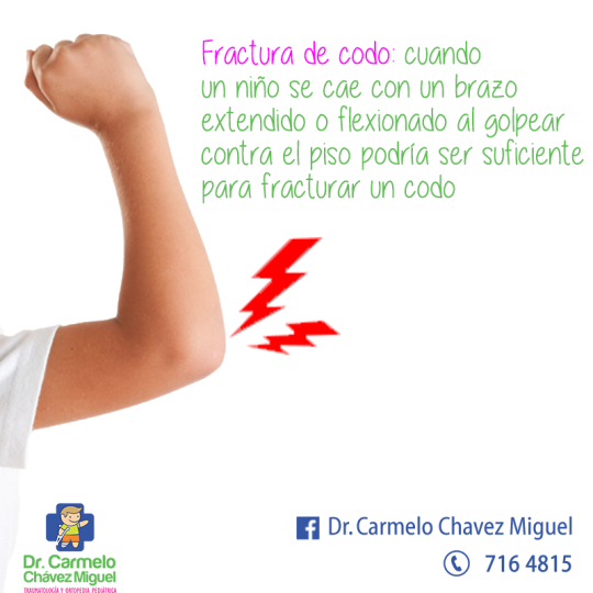
Fractura de codo
Si su niño es un atleta activo o simplemente un niño pequeño que da brincos en su cama, hay grandes probabilidades de que se caiga, en su casa o en el campo de juegos, en algún lugar en cualquier momento.Estas caídas son por lo general inofensivas. Pero cuando un niño se cae con un brazo extendido, la velocidad de la caída combinada con la presión de golpear contra el piso podrían ser suficientes para fracturar, o quebrar, un hueso en el codo. De esa manera ocurren casi todas las fracturas cercanas a la articulación del codo.

Suscribirse a:
Entradas (Atom)


