lunes, 27 de marzo de 2017

Comparación de los resultados de la artroplastia total del hombro reversa, con y sin reparación subscapular

Comparison of reverse total shoulder arthroplasty outcomes with and without subscapularis repair

Fuente
Este artículo es originalmente publicado en:

https://www.ncbi.nlm.nih.gov/pubmed/28277259

http://www.jshoulderelbow.org/article/S1058-2746(16)30448-7/fulltext


De:

Friedman RJ1Flurin PH2Wright TW3Zuckerman JD4Roche CP5.


J Shoulder Elbow Surg. 2017 Apr;26(4):662-668. doi: 10.1016/j.jse.2016.09.027. Epub 2016 Oct 27.


Todos los derechos reservados para:


Copyright © 2017 Journal of Shoulder and Elbow Surgery Board of Trustees. Published by Elsevier Inc. All rights reserved.


Abstract

BACKGROUND:

Repair of the subscapularis with reverse total shoulder arthroplasty (rTSA) is controversial. The purpose of this study is to quantify rTSA outcomes in patients with and without subscapularis repair to determine if there is any impact on clinical outcomes.


CONCLUSIONS:

Significant clinical improvements were observed for both the subscapularis-repaired and non-repaired cohorts, with some statistical differences observed using a variety of outcome measures. Repair of the subscapularis did not lead to inferior clinical outcomes as predicted by biomechanical models. No difference was noted in the complication or scapular notching rates between cohorts. These clinical results show that rTSA using a lateralized humeral prosthesis delivers reliable clinical improvements with a low risk of instability, regardless of subscapularis repair.

KEYWORDS:

Shoulder; arthroplasty; complications; dislocation; reverse; subscapularis


Resumen

ANTECEDENTES:

La reparación del subescapular con artroplastia total reversa del hombro (rTSA) es controvertida. El propósito de este estudio es cuantificar los resultados de rTSA en pacientes con y sin subscapularis reparación para determinar si hay algún impacto en los resultados clínicos.
CONCLUSIONES:Se observaron mejoras clínicas significativas para las cohortes reparadas y no reparadas, con algunas diferencias estadísticas observadas utilizando una variedad de medidas de resultado. La reparación de la subescapularis no condujo a resultados clínicos inferiores como se predice por los modelos biomecánicos. No se observó ninguna diferencia en la complicación o las tasas de muescas escapulares entre cohortes. Estos resultados clínicos muestran que rTSA usando una prótesis humeral lateralizada proporciona mejoras clínicas confiables con un bajo riesgo de inestabilidad, independientemente de la reparación subscapular.


PALABRAS CLAVE:Hombro; Artroplastia; Complicaciones; dislocación; reversa; Subescapular

PMID:   28277259   DOI:   10.1016/j.jse.2016.09.027

Con Técnica de Yesos Seriados y Cirugía de Mínima Invasión, el IMSS Atiende a Bebés con Pie Zambo No.081/2017


El tratamiento inicia en las primeras semanas de vida y continúa hasta los 5 años de edad, cuando el pie está estable.
Los pequeños se benefician con la alineación de los pies y recuperan flexibilidad para realizar sus actividades.
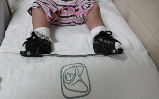
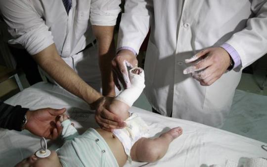
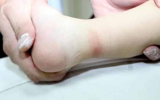
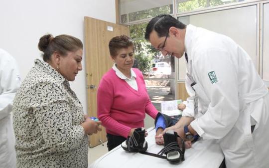
El Servicio de Ortopedia Pediátrica del Hospital “Dr. Victorio de la Fuente Narváez” del IMSS brinda un tratamiento eficaz a bebés que nacen con pie equinovaro aducto congénito (PEVAC), también conocido en Latinoamérica como pie Bott o pie Zambo, mediante la colocación de yesos seriados y una cirugía de mínima invasión, para alinear los pies y recuperar su flexibilidad.
El pie equinovaro es una de las malformaciones congénitas músculo esqueléticas más comunes (torcido hacia adentro, hacia abajo o hacia sí mismo), ocurre en el primer trimestre del desarrollo, se presenta en tres de cada mil nacidos vivos, y afecta a bebés de ambos sexos de manera similar.
El procedimiento médico inicia entre las dos y tres primeras semanas de vida del bebé y se realiza durante las siguientes seis a ocho semanas, con el cambio semanal de yesos para manejar la deformidad que afecta ambos pies.
La técnica de yesos es la opción número uno en el manejo de pie equinovaro con resultados exitosos y se utiliza desde hace más de 50 años a nivel mundial, explicó Juan Agustín Valcarce León, responsable de la Clínica de Yesos del Hospital de Ortopedia de la Unidad Médica de Alta Especialidad “Dr. Victorio de la Fuente Narváez” del Instituto Mexicano del Seguro Social.
Después de la primera fase de tratamiento se evalúa la mejoría de los pies del pequeño para continuar con la cirugía, la cual se aplica en 90 por ciento de los casos. Cosiste en hacer una incisión de dos a tres milímetros en el Tendón de Aquiles, que se regenera y cubre la nueva longitud del pie, agregó el especialista.
En el pasado se hacía una incisión grande en la parte interna del pie, para alargar las estructuras afectadas.
Los beneficios para el paciente son menos tiempo de hospitalización, resultados de buenos a excelentes a largo plazo, en comparación con el método tradicional, en que el paciente sufría más dolor, mayor rigidez y había necesidad de otras cirugías.
El doctor Valcarce León dijo que en la segunda etapa, el niño mantiene la corrección al usar una barra de acero con zapatos unidos a ella durante tres meses, día y noche, y después sólo en las noches, hasta cumplir cinco años de edad, para lograr un pie flexible y no doloroso, que les permite hacer una vida normal, con actividades físicas y deportivas.
Los pacientes continúan en vigilancia médica con una revisión anual hasta los 18 años de edad, generalmente no tienen que usar plantillas ni calzado ortopédico, ya que tienen los pies alineados, flexibles y con fuerza para realizar cualquier actividad.

sábado, 25 de marzo de 2017

Cirugía de ortopedia en el anciano / Orthopedic surgery in the elderly

Marzo 24, 2017. No. 2638


El aumento aislado de troponina cardiaca no modifica el pronóstico de pacientes geriátricos con fractura de cadera
Isolated cardiac troponin rise does not modify the prognosis in elderly patients with hip fracture.
Medicine (Baltimore). 2017 Feb;96(7):e6169. doi: 10.1097/MD.0000000000006169.
Abstract
Perioperative myocardial infarction remains a life-threatening complication in noncardiac surgery and even an isolated troponin rise (ITR) is associated with significant mortality. Our aim was to assess the prognostic value of ITR in elderly patients with hip fracture.... ITR was not associated with a higher risk of new institutionalization or impaired walking ability at 6 months, in contrast to ACS group.In elderly patients with hip fracture, ITR was not associated with a significant increase in death and/or rehospitalization within 6 months.

Artroplastía total de cadera. Seguimiento a 5 años de la evolución funcional pacientes independientes viejos y muy viejos
Total hip arthroplasty for hip fractures: 5-year follow-up of functional outcomes in the oldest independent old and very old patients.
Geriatr Orthop Surg Rehabil. 2014 Mar;5(1):3-8. doi: 10.1177/2151458514520700.
Abstract
INTRODUCTION: This study aimed to determine the dislocation and reoperation rate, functional outcomes, and the survival rate of the unique subset of very old but lucid and independent patients with hip fractures following a total hip arthroplasty (THA) and geriatric team-coordinated perioperative care. METHOD: Between 2000 and 2006, previously independent ambulatory patients ≥80 years old presenting with an intracapsular hip fracture were given THAs under the care of an integrated orthopedic surgery-geriatric service. Their fracture-related complications, ambulation, mental status, and survival were followed for 5 to 11 years postinjury. RESULTS: Five years postinjury, 57 (61.3%) patients of the original study group were living. In all, 3 (3.2%) patients had postoperative hip dislocations (and 2 patients had dislocation twice) and 2 reoperations were needed within the first postoperative month. There were no hip dislocations or reoperations after the first year. Radiographs obtained on 88% of the surviving patients at 5 years postoperatively showed that all remained unchanged from their immediate postoperative images. Nearly half of the patients were still able to ambulate as they did preoperatively and their mixed-model equation was statistically unchanged. CONCLUSION: This study of patients >80 years old with previously good functional status demonstrates that with appropriate surgical (best prosthesis, good operating technique, and regional anesthesia) and geriatric (pre- and postoperative assessments, close follow-up, medication adjustments, and fall-prevention instruction) care, they have few hip dislocations and reoperations, survive postfracture at least as long as their noninjured contemporaries, and continue to function and ambulate as they did prior to their injury.
KEYWORDS: geriatrics; hip fractures; orthogeriatric team; perioperative care; total hip arthroplasty

Mortalidad postoperatoria después de cirugía para fractura de cadera. Seguimiento a tres años
Postoperative Mortality after Hip Fracture Surgery: A 3 Years Follow Up.
PLoS One. 2016 Oct 27;11(10):e0162097. doi: 10.1371/journal.pone.0162097. eCollection 2016.
Abstract
BACKGROUND AND AIMS: To determine mortality rates and predisposing factors in patients operated for a hip fracture in a 3-year follow-up period. METHODS: The study included patients who underwent primary surgery for a hip fracture.The inclusion criteria were traumatic, non-traumatic, osteoporotic and pathological hip fractures requiring surgery in all age groups and both genders. Patients with periprosthetic fractures or previous contralateral hip fracture surgery and patients who could not be contacted by telephone were excluded. At 36 months after surgery, evaluation was made using a structured telephone interview and a detailed examination of the hospital medical records, especially the documents written during anesthesia by the anesthesiologists and the documents written at the time of follow-up visits by the orthopaedic surgeons. A total of 124 cases were analyzed and 4 patients were excluded due to exclusion criteria. The collected data included demographics, type of fracture, co-morbidities, American Society of Anesthesiologists (ASA) scores, anesthesia techniques, operation type (intramedullary nailing or arthroplasty; cemented-noncemented), peroperative complications, refracture during the follow-up period, survival period and mortality causes. RESULTS: The total 120 patients evaluated comprised 74 females(61.7%) and 46 males(38.3%) with a mean age of 76.9±12.8 years (range 23-95 years). The ASA scores were ASA I (0.8%), ASA II (21.7%), ASA III (53.3%) and ASA IV (24.2%). Mortality was seen in 44 patients (36.7%) and 76 patients (63.3%) survived during the 36-month follow-up period. Of the surviving patients, 59.1% were female and 40.9% were male.The survival period ranged between 1-1190 days. The cumulative mortality rate in the first, second and third years were 29.17%, 33.33% and 36.67% respectively. The factors associated with mortality were determined as increasing age, high ASA score, coronary artery disease, congestive heart failure, Alzheimer's disease, Parkinson's disease, malignancycementation and peroperative complications such as hypotension (p<0.05). Mortality was highest in the first month after fracture. CONCLUSION: The results of this study showed higher mortality rates in patients with high ASA scores due to associated co-morbidities such as congestive heart failure, malignancy and Alzheimer's disease or Parkinson's disease. The use of cemented prosthesis was also seen to significantly increase mortality whereas no effect was seen from the anesthesia technique used. Treatment of these patients with a multidiciplinary approach in an orthogeriatric ward is essential. There is a need for further studies concerning cemented vs. uncemented implant use and identification of the best anesthesia technique to decrease mortality rates in these patients.

Curso sobre Anestesia en Trasplantes, Cirugía abdominal, Plástica, Oftalmología y Otorrinolaringología.
Committee for European Education in Anaesthesiology (CEEA) 
y el Colegio de Anestesiólogos de León A.C.
Abril 7-9, 2017, León Guanajuato, México

Informes  (477) 716 06 16, kikinhedz@gmail.com
Congreso Latinoamericano de Anestesia Regional
Asociación Latinoamericana de Anestesia Regional, Capítulo México
Ciudad de México, Mayo 24-27, 2017
Like us on Facebook   Follow us on Twitter   Find us on Google+   View our videos on YouTube 
Anestesiología y Medicina del Dolor

52 664 6848905

Copyright © 2015

Lesiones atléticas de la cadera


Lesiones atléticas de la cadera » Athletic Hip Injuries

Fuente
Este artículo es originalmente publicado en:

https://www.ncbi.nlm.nih.gov/pubmed/28252476
http://journals.lww.com/jaaos/Fulltext/2017/04000/Athletic_Hip_Injuries.3.aspx

De:
Lynch TS1,Bedi A,Larson CM.
J Am Acad Orthop Surg.
2017 Apr;25(4):269-279. doi: 10.5435/JAAOS-D-16-00171.

Todos los derechos reservados para:

© 2017 by American Academy of Orthopaedic Surgeons


Abstract
Historically, athletic hip injuries have garnered little attention; however, these injuries account for approximately 6% of all sports injuries and their prevalence is increasing. At times, the diagnosis and management of hip injuries can be challenging and elusive for the team physician. Hip injuries are seen in high-level athletes who participate in cutting and pivoting sports that require rapid acceleration and deceleration. Described previously as the “sports hip triad,” these injuries consist of adductor strains, osteitis pubis, athletic pubalgia, or core muscle injury, often with underlying range-of-motion limitations secondary to femoroacetabular impingement. These disorders can happen in isolation but frequently occur in combination. To add to the diagnostic challenge, numerous intra-articular disorders and extra-articular soft-tissue restraints about the hip can serve as pain generators, in addition to referred pain from the lumbar spine, bowel, bladder, and reproductive organs. Athletic hip conditions can be debilitating and often require a timely diagnosis to provide appropriate intervention.


Resumen
Históricamente, las lesiones atléticas de la cadera han ganado poca atención; Sin embargo, estas lesiones representan aproximadamente el 6% de todas las lesiones deportivas y su prevalencia está aumentando. A veces, el diagnóstico y el manejo de las lesiones de cadera puede ser difícil y elusivo para el médico del equipo. Las lesiones de cadera se observan en atletas de alto nivel que participan en deportes de corte y pivotantes que requieren una rápida aceleración y desaceleración. Descrita anteriormente como la “tríada de la cadera deportiva”, estas lesiones consisten en cepas de aductores, osteítis púbico, pubalgia atlética o lesión muscular básica, a menudo con limitaciones subyacentes del rango de movimiento secundarias al choque femoroacetabular. Estos trastornos pueden ocurrir en el aislamiento pero ocurren con frecuencia en combinación. Para añadir al reto diagnóstico, numerosos trastornos intra articulares y restricciones extra articulares de tejidos blandos alrededor de la cadera pueden servir como generadores de dolor, además del dolor referido desde la columna lumbar, el intestino, la vejiga y los órganos reproductivos. Las condiciones atléticas de la cadera pueden ser debilitantes ya menudo requieren un diagnóstico oportuno para proporcionar la intervención apropiada.


PMID: 28252476 DOI:
10.5435/JAAOS-D-16-00171

#lesiones deportivas #lesiones de cadera #atletas #cadera