jueves, 8 de junio de 2017

Hipertermia maligna / Malignant hyperthermia

Junio 8, 2017. No. 2713


Estimad@ Dr@ Víctor Valdés:  


Chichen Itza

Rabdomiólisis por ejercicio  y golpe de calor: Tenga cuidado con la sedación volátil anestésica.
Exertional rhabdomyolysis and heat stroke: Beware of volatile anesthetic sedation.
World J Crit Care Med. 2017 Feb 4;6(1):21-27. doi: 10.5492/wjccm.v6.i1.21. eCollection 2017 Feb 4.
Abstract
In view of the enormous popularity of mass sporting events such as half-marathons, the number of patients with exertional rhabdomyolysis or exercise-induced heat stroke admitted to intensive care units (ICUs) has increased over the last decade. Because these patients have been reported to be at risk for malignant hyperthermia during general anesthesia, the intensive care community should bear in mind that the same risk of life-threatening rhabdomyolysis is present when these patients are admitted to an ICU, and volatile anesthetic sedation is chosen as the sedative technique. As illustrated by the three case studies we elaborate upon, a thorough diagnostic work-up is needed to clarify the subsequent risk of strenuous exercise, and the anesthetic exposure to volatile agents in these patients and their families. Other contraindications for the use of volatile intensive care sedation consist of known malignant hyperthermia susceptibility, congenital myopathies, Duchenne muscular dystrophy, and intracranial hypertension.
KEYWORDS: Congenital myopathies; Exertional rhabdomyolysis; Heat stroke; Inhalational anesthetics; Intensive care sedation; Malignant hyperthermia

Hipertermia  maligna. Una revisión
Malignant hyperthermia: a review.
Orphanet J Rare Dis. 2015 Aug 4;10:93. doi: 10.1186/s13023-015-0310-1.
Abstract
Malignant hyperthermia (MH) is a pharmacogenetic disorder of skeletal muscle that presents as a hypermetabolic response to potent volatile anesthetic gases such as halothane, sevoflurane, desflurane, isoflurane and the depolarizing muscle relaxant succinylcholine, and rarely, in humans, to stressors such as vigorous exercise and heat. The incidence of MH reactions ranges from 1:10,000 to 1: 250,000 anesthetics. However, the prevalence of the genetic abnormalities may be as great as one in 400 individuals. MH affects humans, certain pig breeds, dogs and horses. The classic signs of MH include hyperthermia, tachycardia, tachypnea, increased carbon dioxide production, increased oxygen consumption, acidosis, hyperkalaemia, muscle rigidity, and rhabdomyolysis, all related to a hypermetabolic response. The syndrome is likely to be fatal if untreated. An increase in end-tidal carbon dioxide despite increased minute ventilation provides an early diagnostic clue. In humans the syndrome is inherited in an autosomal dominant pattern, while in pigs it is autosomal recessive. Uncontrolled rise of myoplasmic calcium, which activates biochemical processes related to muscle activation leads to the pathophysiologic changes. In most cases, the syndrome is caused by a defect in the ryanodine receptor. Over 400 variants have been identified in the RYR1 gene located on chromosome 19q13.1, and at least 34 are causal for MH. Less than 1 % of variants have been found in CACNA1S but not all of these are causal. Diagnostic testing involves the in vitro contracture response of biopsied muscle to halothane, caffeine, and in some centres ryanodine and 4-chloro-m-cresol. Elucidation of the genetic changes has led to the introduction of DNA testing for susceptibility to MH. Dantrolene sodium is a specific antagonist and should be available wherever general anesthesia is administered. Increased understanding of the clinical manifestation and pathophysiology of the syndrome, has lead to the mortality decreasing from 80 % thirty years ago to <5 % in 2006.

Manejo de la hipertermia maligna. Diagnóstico y tratamiento
Management of malignant hyperthermia: diagnosis and treatment.
Ther Clin Risk Manag. 2014 May 14;10:355-62. doi: 10.2147/TCRM.S47632. eCollection 2014.
Abstract
Malignant hyperthermia is a potentially lethal inherited disorder characterized by disturbance of calcium homeostasis in skeletal muscle. Volatile anesthetics and/or the depolarizing muscle relaxant succinylcholine may induce this hypermetabolic muscular syndrome due to uncontrolled sarcoplasmic calcium release via functionally altered calcium release receptors, resulting in hypoxemia, hypercapnia, tachycardia, muscular rigidity, acidosis, hyperkalemia, and hyperthermia in susceptible individuals. Since the clinical presentation of malignant hyperthermia is highly variable, survival of affected patients depends largely on early recognition of the symptoms characteristic of malignant hyperthermia, and immediate action on the part of the attending anesthesiologist. Clinical symptoms of malignant hyperthermia, diagnostic criteria, and current therapeutic guidelines, as well as adequate management of anesthesia in patients susceptible to malignant hyperthermia, are discussed in this review.
KEYWORDS: genetics; in vitro contracture test; malignant hyperthermia; succinylcholine; volatile anesthetics

Hipertermia maligna
Malignant hyperthermia.
Swiss Med Wkly. 2012 Jul 31;142:w13652. doi: 10.4414/smw.2012.13652.
Abstract
Malignant hyperthermia (MH) is a subclinical myopathy, usually triggered by volatile anaesthetics and depolarising muscle relaxants. Clinical symptoms are variable, and the condition is sometimes difficult to identify. Nevertheless, rapid recognition and specific as well as symptomatic treatment are crucial to avoid a lethal outcome. Molecular genetic investigations have confirmed the skeletal muscle type ryanodine receptor to be the major MH locus with more than 70% of MH families carrying a mutation in this gene. There is no screening method to test for MH, as current tests are invasive (open muscle biopsy) or restricted to MH families with known MH-associated mutations (molecular genetic testing). The prevalence of the MH trait is unknown, because the clinical penetrance after contact with triggering agents is very variable. More recently, MH mutations have been associated with rhabdomyolysis following statin therapy or with non-pharmacological triggering, such as exertional heat stroke.


IX Foro Internacional de Medicina del Dolor y Paliativa
Instituto Nacional de Ciencias Médicas y Nutrición Salvador Zubirán
Ciudad de México, 8 al 10 de Junio
Like us on Facebook   Follow us on Twitter   Find us on Google+   View our videos on YouTube 
Anestesiología y Medicina del Dolor

52 664 6848905

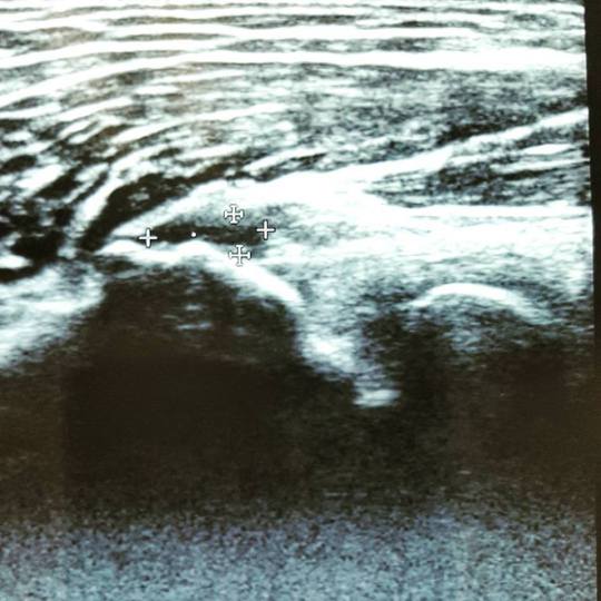
Ruptura parcial aguda del tendón extensor común del codo


Acute partial rupture of the common extensor tendon

Fuente
Este artículo es originalmente publicado en:
De:
2010 Jun;13(2):74-5. doi: 10.1016/j.jus.2010.07.002. Epub 2010 Aug 3.
Todos los derechos reservados para:

Author information ► Copyright and License information ▼

Copyright © 2010 Elsevier Masson Italy.


Abstract
in English , Italian
Rupture of the common extensor tendon is the most common acute tendon injury of the elbow. The authors describe a case of a patient with a clinical history of tendinopathy caused by functional overload of the common extensor tendon, treated also with infiltrations of steroids, and subsequent partial rupture of the tendon during sport activity. The diagnosis was made clinically and at ultrasound (US) examination; US follow-up after some time showed the healing of the lesion. This case confirms that injections of steroids may be a contributory cause of tendon rupture, and emphasizes the sensitivity and specificity of US in the study of pathologies of the elbow tendons.
KEYWORDS:
Common extensor tendon; Elbow; Ultrasonography
Resumen
En inglés, italiano
La ruptura del tendón extensor común es la lesión del tendón agudo más común del codo. Los autores describen un caso de un paciente con antecedentes clínicos de tendinopatía por sobrecarga funcional del tendón extensor común, tratado también con infiltraciones de esteroides y posterior ruptura parcial del tendón durante la actividad deportiva. El diagnóstico se realizó clínicamente y con ecografía (US); El seguimiento de EE.UU. después de algún tiempo mostró la curación de la lesión. Este caso confirma que las inyecciones de esteroides pueden ser una causa contribuyente de la ruptura del tendón, y hace hincapié en la sensibilidad y especificidad de EE.UU. en el estudio de las patologías de los tendones del codo.
PALABRAS CLAVE:
Tendón extensor común; Codo; Ultrasonografía
PMID:23396798      PMCID:
DOI:

IV Congreso Internacional de la Sociedad de Cirugía de Acetábulo y Pelvis


 a llevarse a cabo los días 7, 8 y 9 de septiembre en SLP, México.
Registro e inscripciones en:

miércoles, 7 de junio de 2017

OPTIMIZACIÓN DE ANALGÉSICOS PARA UNA MAYOR PARTICIPACIÓN EN LA TERAPIA DE EJERCICIO ENTRE PACIENTES CON OSTEOARTRITIS DE RODILLA Y DOLOR SEVERO: UN ESTUDIO DE FACTIBILIDAD.


Optimization of Analgesics for Greater Exercise Therapy Participation Among Patients With Knee Osteoarthritis and Severe Pain: A Feasibility Study.

Fuente
Este artículo es originalmente publicado en:
De:
2016 Mar;68(3):332-40. doi: 10.1002/acr.22682.
Todos los derechos reservados para:

© 2016, American College of Rheumatology.


Abstract
OBJECTIVE:
Severe pain in patients with knee osteoarthritis (OA) hampers the ability to exercise. A protocol for the standardized optimization of analgesics in combination with exercise therapy was developed. The purpose of this protocol was to reduce pain, thereby allowing the patient to participate in exercise therapy. The objective of the present study was to evaluate the feasibility and outcome of the protocol.
CONCLUSION:
The combined intervention of standardized analgesic prescription and exercise therapy allows most patients with knee OA and severe pain to participate in exercise therapy, leading to reduction of pain and activity limitations. These promising results need to be confirmed in a randomized controlled trial.
Resumen
OBJETIVO:
El dolor severo en pacientes con osteoartritis de rodilla (OA) dificulta la capacidad de hacer ejercicio. Se desarrolló un protocolo para la optimización estandarizada de los analgésicos en combinación con la terapia con ejercicios. El propósito de este protocolo fue reducir el dolor, permitiendo al paciente participar en la terapia de ejercicios. El objetivo del presente estudio fue evaluar la viabilidad y los resultados del protocolo.
CONCLUSIÓN:
La intervención combinada de prescripción analgésica estandarizada y terapia de ejercicios permite a la mayoría de los pacientes con OA de rodilla y dolor intenso participar en terapia de ejercicios, lo que lleva a la reducción del dolor y las limitaciones de actividad. Estos resultados prometedores deben confirmarse en un ensayo controlado aleatorio.
© 2016, American College of Rheumatology.
PMID: 26239403   DOI:
[Indexed for MEDLINE]
Free full text